Photo gallery: Dental corner: Assisting a cat with a jaw fracture
See how a veterinary team works together to help a cat suffering from a mandible fracture after a car accident.
Next >

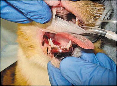
A 7-year-old neutered male domestic shorthaired cat was presented to the emergency service of the University of Pennsylvania's School of Veterinary Medicine after being hit by a car. He was treated for shock and sent for chest radiographs. Luckily, he only sustained head trauma. Figure 1 shows the symphyseal separation, the gap between the cat's mandibular incisors, and the sublingual bruising.
Photos courtesy of the University of Pennsylvania
Click the Next button to go the next page.

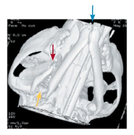
The Dentistry and Oral Surgery Service was called in to evaluate his injuries. He was anesthetized and underwent a CT scan, which showed a symphyseal separation and right mandible fractures in two places. An oral examination revealed some minor gingival and sublingual bruising, but no fractured teeth. In figure 2, the blue arrow shows the symphyseal separation, and the red and yellow arrows show the mandibular fractures.
Click the Next button to go the next page.

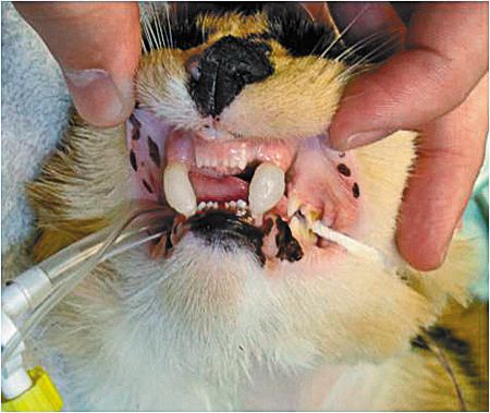
To stabilize the jaw fractures, the cat's canine teeth were bonded together with an acrylic material to fixate the jaws in position until the bones healed. This technique ensures no movement of the unstable mandible while still allowing for some tongue movement. An esophagostomy tube was placed to ensure the cat's nutritional needs were met, and a fentanyl patch was placed on his lateral chest area. Figure 3 shows the acrylic bonding of the canine teeth.
Click the Next button to go the next page.

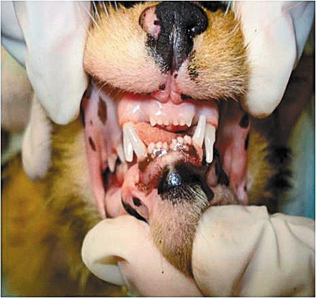
At his two-week recheck appointment, the cat was eating on his own, while being supplemented through his esophagostomy tube. At eight weeks, he was anesthetized again and the acrylic material was removed from the canine teeth with a high-speed handpiece and extraction forceps. Intraoral radiographs showed that the fracture sites had healed and that the jaw occlusion was good. The esophagostomy tube was removed, and the cat recovered nicely. The technician's role is varied, from assisting with radiographs and surgery to assisting with feeding tube placement. The technician also plays a role in education and after care. Figure 4 shows the post-removal of the acrylic bonding to see how the jaws align now.
Click the Next button to go the beginning.