Pulmonary edema (Proceedings)
Pulmonary edema is defined as the accumulation of abnormal amount of extravascular lung water. Pulmonary edema may range from clinically insignificant to life-threatening. Pulmonary edema forms when there is an alternation in the balance of Starling forces (hydrostatic and colloid osmotic) between the interstitium and pulmonary capillary beds that favors increasing filtration to the interstitium. Increased capillary hydrostatic forces will result in a low protein edema fluid while lowered colloid osmotic forces will promote a high protein edema fluid.
Definition and Classification
Pulmonary edema is defined as the accumulation of abnormal amount of extravascular lung water. Pulmonary edema may range from clinically insignificant to life-threatening. Pulmonary edema forms when there is an alternation in the balance of Starling forces (hydrostatic and colloid osmotic) between the interstitium and pulmonary capillary beds that favors increasing filtration to the interstitium. Increased capillary hydrostatic forces will result in a low protein edema fluid while lowered colloid osmotic forces will promote a high protein edema fluid.
Pulmonary edema is generally divided into cardiogenic and non-cardiogenic forms based upon the etiology. Cardiogenic pulmonary edema results from increased pulmonary capillary hydrostatic pressure caused by left-sided heart failure. Common causes of left-sided heart failure in small animals include dilated cardiomyopathy, acquired mitral valve regurgitation, and hypertrophic cardiomyopathy. Non-cardiogenic edema results from decreased colloid osmotic pressure or altered vascular permeability in the pulmonary capillaries. Common veterinary causes of non-cardiogenic pulmonary edema include upper airway obstruction (transient), electric cord injury, and sepsis/acute respiratory distress syndrome (ARDS).
History/Symptoms
In many dogs with cardiogenic pulmonary edema there is a history of a heart murmur due to endocardiosis. Other historical complaints for cardiogenic pulmonary edema include tachypnea or orthopnea, respiratory distress and coughing. Cats with heart failure may have had no premonitory signs other than acute onset of respiratory distress.
In animals with non-cardiogenic pulmonary edema, they may have had an event, that is a known trigger of non-cardiogenic pulmonary edema (i.e. electric cord injury, upper airway obstruction or seizures) or the patient may be hospitalized with serious illness. In either case, signs of pulmonary edema include tachypnea and respiratory distress.
Risk factors for the development of cardiogenic pulmonary edema include heart disease, high sodium meals and overzealous fluid therapy (crystalloid or colloid).Risk factors for the development of non-cardiogenic edema include accidental exposure to a trigger or severe underlying illness.
Physical Signs
Physical signs of cardiogenic edema include tachypnea and respiratory distress.
Auscultation may reveal pulmonary crackles or the presence of cardiac abnormalities such as a murmur, gallop or arrhythmia. Heart rate is usually rapid and pulse quality weak. Jugular vein distension or ascites may be present in cases of biventricular heart failure. Dogs with long-standing cardiac disease are often thin to emaciated (cardiac cachexia). Some cats may have enlarged thyroid gland and almost all cats with cardiogenic pulmonary edema are hypothermic. Physical signs of non-cardiogenic edema include tachypnea and respiratory distress. Auscultation may reveal crackles but heart sound and rate are generally normal. Animals with electric cord injury may have burns on the tongue, palate or oral commissures. Animals with upper airway obstruction may have loud or noisy breathing (laryngeal paralysis/brachycephalic airway syndrome). Animals with systemic illness may show an acute or rapidly progressive increase in respiratory rate and effort.
Diagnostic Testing
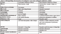
In all forms of pulmonary edema, the diagnostic test of choice is thoracic radiographs. Thoracic radiographs will demonstrate interstitial to alveolar infiltrates. Distribution of infiltrates may aid in identification of the etiology. In human medicine, pulmonary capillary wedge pressure (PCWP) is used as the gold standard to distinguish cardiogenic from non-cardiogenic pulmonary edema. An elevated PCWP (>18 mmHg) is diagnostic of cardiac dysfunction. The measurement of PCWP requires the placement of a pulmonary artery catheter and is not routinely performed in veterinary medicine at this point. Attempts to substitute central venous pressure for PCWP have not been overwhelmingly worthwhile.
In dogs with cardiogenic pulmonary edema, thoracic radiographs will usually document cardiomegaly, pulmonary venous distension and interstitial to alveolar infiltrates. In dogs, the infiltrates generally begin in the perihilar region, but may expand to fill the entire parenchyma in severe cases. In cats with cardiogenic edema, thoracic radiographs will often document cardiomegaly and pulmonary venous distension. The pattern of pulmonary edema in cats with heart failure may be variable in distribution. Other diagnostic tests for cardiogenic pulmonary edema include echocardiography and EKG analysis and an elevated PCWP.
In animals with non-cardiogenic edema, thoracic radiographs will show pulmonary infiltrates without cardiomegaly. The classic distribution of non-cardiogenic pulmonary edema from upper airway obstruction, electric cord injury or seizures is in the dorsal caudal lung fields. Non-cardiogenic pulmonary edema from other causes will be distributed through the lung fields. Other diagnostic tests for non-cardiogenic pulmonary edema include a normal echocardiogram and normal pulmonary capillary wedge pressure.
Measurement of the ratio of the protein content of the pulmonary edema fluid to the protein content of the plasma is also useful to determine the etiology (cardiogenic Vs non-cardiogenic) of the edema. Cardiogenic edema fluid is low protein and the ratio is <0.5 (usually 0.3) and in non-cardiogenic edema the protein is high and the ratio >0.5 (usually 0.7-0.8). It is may be difficult to collect sample of edema fluid unless the animal is intubated or coughing up edema fluid.
Treatment
Treatment of cardiogenic pulmonary edema centers on oxygen, rest, diuretics and vasodilators. The most commonly used diuretic is furosemide although others such as spironolactone or hydrochlorothiazide may be used with more chronic heart failure. Vasodilators include nitroglycerine, nitroprusside, ACE inhibitors (enalapril) or hydralazine. Vasodilators should be used with extreme caution (if at all) in animals with systemic hypotension. Animals should be allowed to rest and respiratory rate/effort, heart rate and blood pressure should be monitored. In dogs with dilated cardiomyopathy, positive inotropic support with dobutamine may be provided. Significant tachyarrhythmias may be addressed with anti-arrhythmic therapy as needed. Rarely, mechanical ventilation may be provided. Intravenous Fluids Are Contraindicted At Any Rate In An Animal With Acute Congestive Heart Failure, Although Animals Should Be Offered Water. The prognosis for acute CHF is fair, although animals will require life-long cardiac therapy and their long-term prognosis is guarded.
Treatment of animals with non-cardiogenic pulmonary edema is less straightforward than treatment of cardiogenic pulmonary edema. Animals with acute non-cardiogenic edema from electric cord injury, upper airway obstruction generally improve rapidly if allowed to rest in oxygen. Some clinicians advocate therapy diuretics, anti-inflammatories (i.e. dexamethasone) or colloids (i.e. hetastarch) although no compelling evidence exists to favor one form of therapy over another. In animals with non-cardiogenic pulmonary edema from other sources, therapy is dependent upon the underlying cause, but may include antibiotics or intravenous fluids. Dogs with severe non-cardiogenic pulmonary edema may occasionally require mechanical ventilatory support. The prognosis for non-cardiogenic edema ranges from good to grave.
In all forms of pulmonary edema, monitoring should include respiratory rate and effort, heart rate, blood pressure, body temperature and oxygen saturation (pulse oximetry/arterial blood gas analysis). The veterinarian should remember to let the patient rest in a stress-free environment as much as possible.
Patients with pulmonary edema may be challenging to manage. Early attempts should be made to distinguish cardiogenic from non-cardiogenic pulmonary edema. The prognosis is dependent upon the underlying cause.

Chart 1. Distinguishing characteristics of cardiogenic versus non-cardiogenic pulmonary edema.
A guide for assessing respiratory emergencies
November 15th 2024Mariana Pardo, BVSc, MV, DACVECC, provided an overview on breathing patterns, respiratory sounds, lung auscultation; and what these different sounds, patterns, and signs may mean—and more—in her lecture at the 2024 NY Vet Show
Read More