Pyothorax in cats and dogs (Proceedings)
The purpose of this lecture is to review the management of pyothorax in cats and dogs. Pulmonary infection can result from bacterial, viral, fungal or protozoal infection, however, pyothorax is almost uniformly bacterial. The pleural space has a small amount of fluid normally (~ 5 ml) which serves as lubrication for the pulmonary parenchyma.
The purpose of this lecture is to review the management of pyothorax in cats and dogs. Pulmonary infection can result from bacterial, viral, fungal or protozoal infection, however, pyothorax is almost uniformly bacterial. The pleural space has a small amount of fluid normally (~ 5 ml) which serves as lubrication for the pulmonary parenchyma. However, liters of fluid cross the pleural space daily through extravascation and subsequent resorption. This is important as inflammation and loss of resorptive ability can result in large volume of effusion accumulating quickly. Pyothorax is defined as a septic pleural effusion. In people the term empyema is often used. The organisms can be a mixed population including anaerobic organisms or can be a single organism depending on the initial sources of the infection.
Diagnosis is made by cytological identification of bacterial organisms in pleural effusion, and ideally by positive bacterial culture and sensitivity results. Cytological examination should also document supparative inflammation with toxic change and intracellular bacteria. In dogs and cats, most commonly pyothorax is a mixed population of bacteria, and the fluid is clearly purulent in appearance. This photograph shows the classical "tomato soup" exudate that is common in canine pyothorax. In people, as empyema is often less apparently infected, complementary fluid analysis, including evaluation such as glucose, LDH or amylase are occasionally employed.

Risk factors for the development of pyothorax in dogs include large breed young active hunting dogs. Pyothorax is through to develop in response to migration of the inhaled plant awns, and are often associated with pulmonary abscesses. In cats, pyothorax is more common in multiple cat households, and in those cats that go outside. The most common cause of feline pyothorax are thought to include thoracic bite wounds and extension from pulmonary abscesses (eg. Severe URI/pneumonia). Pyothorax in both cats and dogs can be associated with esophageal perforation or with iatrogenic infection associated with thoracocentesis or thoracotomy.
Complementary diagnostics to cytology and bacterial culture include CBC/Chemistry profile/UA as well as Feline leukemia/FIV testing. White blood counts are often markedly elevated in dogs and cats, and a profound left shift is often present. Chemistry profiles are typically normal, but may document low albumin or evidence of other systemic disease (azotemia, raised liver enzymes). FeLV or FIV could affect the ability of the cat respond to the supportive care, or at least in the case of FeLV, might affect the client's wishes as far as proceeding with intensive supportive care. From an imaging standpoint, radiographs will usually simply support the presence of effusion, although mass lesion or spontaneous pneumothorax may also be present. Ultrasound examination or computed tomography could be considered for more careful examination of the thoracic cavity, in particular for a search for an abscess or foreign body. Recall that the mediastinum in both cats and dogs is not intact, thus unilateral effusions reflect the presence of fibrosis adhesions occluding the exchange of fluid across the mediastinum. For similar reasons, the long standing practice of recording the volumes that are obtained from the right and left sides of the chest is unlikely helpful, as the fluid can easily move from one side to the other.
Following diagnosis, the major questions that exist include 1)prognosis and 2) best approach to therapy. Prognosis can be divided into short term survival and long term rates of recurrence or on-going pulmonary compromise. Prognosis, like in most cases of sepsis, will reflect the degree of systemic inflammation and multiple organ dysfunction. Overall, pyothorax tends to have a better prognosis that other causes of pleural effusion, in almost all cases successful therapy is extensive and may require significant financial commitment on behalf of the client and a longer duration of hospitalization that more many other diseases. Cats that present moribund (or interestingly with pytalism) have a worse prognosis. Long term prognosis can be more variable, as recurrence is not uncommon, especially in dogs with Actinomyces infection or potentially in animals treated more conservatively.
The options for therapy include medical or surgical therapy. All forms of therapy include an extended course of broad spectrum antibiotics, ideally directed by culture and sensitivity testing. Animals should ideally be hospitalized so they can be more closely monitored and receive 24 hours care. Other therapies include discussion of the following:
Medical therapy
• Periodic thoracocentesis
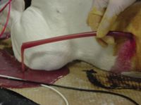
• Chest tube placement; chest tube can be placed on one side or bilaterally. Most commonly, they are placed bilaterally. Chest tube are typically large bore, but a small bore chest tube (MilaInternational) has been used effectively in recent years. These tubes are easy to place, and have been well-tolerated by most animals.
o Intermittent hand aspiration; In this case, the tube is aspirated every 4 hours and the volume recorded.
o Aspiration with saline or heparinzed saline flushing; this technique relies upon infusing 10-20 ml/kg into the thorax and then re-aspirating the fluid. Heparin is added at 10-20 units/ml, with the goal of decreasing adhesions.
o Aspiration with infusion of antibiotics- some clinicians tote this technique, however, due to the vascular nature of the pleural space it is unlikely any benefit over systemic antibioics.
o Aspiration with infusion of local thrombolytics is occasionally pursed in people with extensive pleural adhesions. Management of a horse with pleuropneumonia with thrombolytics has been described.
Surgical therapy (delayed or immediate)
• Median sternotomy
• Lateral thoracotomy
The right approach is often hotly debated. One study (Rooney et al) has shown an improved outcome in dogs treated with surgical intervention in comparison to dogs treated medically. Based upon this retrospective study, as well as personal experience, the recommendation is to explore dogs via a median sternotomy as soon as practical (eg, not at 2 am) rather than first placing chest tubes and treating more conservatively first. However, a more recent retrospective (Boothe et al) showed no improvement with surgical management, but advocated lavage with heparin. In cats, most often the recommendations have been to treat medically for 4-7 days first, and then if there is no improvement, to consider surgical exploration at that point. Surgical exploration should be pursued more promptly if there is evidence of a pulmonary abscess or other lesion that appears to be surgical resectable.
References
Evaluation of outcomes in dogs treated for pyothorax: 46 cases (1983-2001).
Boothe HW, Howe LM, Boothe DM, Reynolds LA, Carpenter M.J Am Vet Med Assoc. 2010 Mar 15;236(6):657-63.
Evaluation of small-bore wire-guided chest drains for management of pleural space disease.
Valtolina C, Adamantos S.J Small Anim Pract. 2009 Jun;50(6):290-7.
Septic pneumonia and pyothorax in dogs.
Lewis DH, Armitage-Chan E, Chan DL, Garden O.Vet Rec. 2009 Mar 21;164(12):378. No abstract available.
Medical and surgical management of pyothorax.
MacPhail CM.Vet Clin North Am Small Anim Pract. 2007 Sep;37(5):975-88, vii. Review.
Risk factors, prognostic indicators, and outcome of pyothorax in cats: 80 cases (1986-1999).
Waddell LS, Brady CA, Drobatz KJ.J Am Vet Med Assoc. 2002 Sep 15;221(6):819-24.
Bacteria associated with pyothorax of dogs and cats: 98 cases (1989-1998).
Walker AL, Jang SS, Hirsh DC.J Am Vet Med Assoc. 2000 Feb 1;216(3):359-63.
A guide for assessing respiratory emergencies
November 15th 2024Mariana Pardo, BVSc, MV, DACVECC, provided an overview on breathing patterns, respiratory sounds, lung auscultation; and what these different sounds, patterns, and signs may mean—and more—in her lecture at the 2024 NY Vet Show
Read More