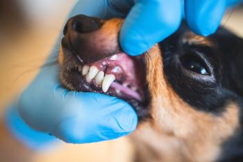
Recurrent oral hemorrhage in a cat – an unusual etiology
Spontaneous oral hemorrhage without trauma is an uncommon finding.
Oral hemorrhage is an uncommon clinical finding when it occurs spontaneously without a history of trauma. Nontraumatic oral bleeding can be due to neoplasia, periodontal disease and, in the case discussed below, what appears to be several factors. Frank and significant hemorrhage as seen in this case is particularly unusual.
A curious case
A white domestic shorthaired cat was presented to a local emergency facility for evaluation of spontaneous oral bleeding. No abnormalities other than frank oral hemorrhage were noted on presentation. The patient was sedated, and an oral examination revealed arterial hemorrhage associated with a diffuse palatal defect 3 mm palatal to the right maxillary canine tooth. A suture was placed into the palatal tissue and successfully stopped the hemorrhage. The patient was kept for observation and then discharged some time later.
Less than 48 hours later, the patient was presented again to the emergency facility with the same complaint. Sedation and suturing were repeated at the same site, although this time a horizontal mattress suture was placed. A recommendation was made for referral the next morning to determine a cause for the recurrent spontaneous hemorrhage.
Photo 1: Three palatal lesions are evident. The lesion adjacent to the canine tooth was the origin of the bleeding. (Photos: Courtesy of Dr. Brett Beckman)
On physical examination at the referral clinic, numerous fleas, pink staining of the coat in multiple locations about the torso and limbs and dried blood within and about the oral cavity were noted. The owners were queried and confirmed that the cat groomed excessively, but they were not aware of the flea infestation. The results of a complete blood count, serum chemistry profile, T4 concentration measurement and feline leukemia virus and feline immunodeficiency virus testing revealed a slight anemia (PCV = 23.7%), mild hypoglobulinemia and a mild decrease in the T4 concentration. While awaiting sedation and a complete oral examination, the patient's oral hemorrhage returned, with arterial blood dripping freely from the oral cavity.
Photo 2: A palatal mucosal flap was used to visualize the bone adjacent to the defect, ligate the major palatine artery and obtain biopsy samples of the lesion.
The patient was sedated, and an oral examination revealed bleeding associated with the palatal lesion. The tissue associated with the lesion was dark-red and lacked palatal ruggae (Photo 1). Two additional smooth darkened lesions were present in the caudal palatal mucosa.
Photo 3: The major palatine artery is seen here after surgical exposure.
Because of the recurrence and severity of the hemorrhage, ligation of the major palatine artery was indicated to eliminate the major arterial supply to the tissue. A full-thickness palatal flap was used to expose the bone adjacent to the lesion for visualization and to isolate the artery for ligation (Photos 2 and 3). The bone and adjacent palatal tissue appeared normal. Biopsy samples were then taken of the bleeding lesion and one of the caudal palatal mucosal lesions. The flap was closed by using a simple interrupted suture technique (Photo 4). Eosinophilic granuloma and neoplasia were considered the most likely rule-outs. Histopathology was nonspecific and revealed well-differentiated hyperplastic epithelial cells. Pruritic dermatitis secondary to flea infestation was diagnosed based on the clinical signs and was resolved with flea treatment and an anti-inflammatory dose of prednisone given every other day.
Photo 4: The flap is sutured after ligation.
The patient returned in six weeks for a recheck. No additional bleeding was observed, and external parasites were absent. Evidence of all three lesions was still present, but the lesions appeared to be resolving and not clinically active (Photo 5).
Photo 5: The patient at a recheck showing the resolving lesions.
Discussion
Definitive evidence of the association between the bleeding and grooming behavior was suspected but could not be confirmed. However, it is likely that self-induced trauma to the palate, associated with excessive grooming, played a role in the origination and persistence of the recurring hemorrhage in this case. Personal communication with a colleague from New Zealand1 who has experienced these cases on several occasions suggested that the hair becomes very rough due to grooming and increases palatal trauma. Ligation of the major palatine artery and external parasite control were curative in those cases. The lack of ruggae may have been due to persistent abrasion from the dorsum of the tongue. No tongue lesions were observed.
No published reports of these cases exist, but the 2007 Veterinary Dental Forum Proceedings included reports of two similar cases in Japan.2 External parasites and excessive grooming were not mentioned in these reports. Both cats were retrovirus-positive, unlike in this case, but had lesions in an identical location to those described here. In addition, one cat had a low T4 concentration. Histopathology results were identical in each of the cats from Japan, demonstrating erosive and chronic inflammation with lymphocytic and mastocytic infiltration. Additionally, rich vascularization was observed at the submucosa.
Multiple factors are likely involved in these cases. Speculation based on this current case and past cases points to an unusual etiology, although definitive answers can only be drawn with additional cases and documentation. I encourage readers who have documentation — preferably with images and histopathology — associated with similar cases to the one described here to contact me via e-mail at
Dr. Beckman is acting president of the American Veterinary Dental Society and owns and operates a companion-animal and referral dentistry and oral surgery practice in Punta Gorda, Fla. He sees referrals at Affi liated Veterinary Specialists in Orlando and at Georgia Veterinary Specialists in Atlanta, lectures internationally and operates the Veterinary Dental Education Center in Punta Gorda.
References
1. Russel Tucker, DVM, DAVDC. Personal communication, Kohimarama, Auckland, New Zealand, 1071.
2. Eguchi, Chimura, S, Okuda A. Surgical correction of continual bleeding from ulcerative lesions on the hard palate of 2 cat cases, in Proceedings. Vet Dental Forum 2007.
Newsletter
From exam room tips to practice management insights, get trusted veterinary news delivered straight to your inbox—subscribe to dvm360.


