Respiratory disease caused by parasites (Proceedings)
Parasites are major causes of respiratory tract disease in the dog and cat. Recent advances in therapy of these diseases have been made providing the practicing veterinarian with a more rational treatment modality.
Parasites are major causes of respiratory tract disease in the dog and cat. Recent advances in therapy of these diseases have been made providing the practicing veterinarian with a more rational treatment modality. This review will discuss the biology, diagnosis, disease, and treatment of respiratory parasites (protozoan, nematode, trematode, and arthropods) of the dog and cat. Emphasis will be placed on the use of modern chemotherapeutic agents in their control. The parasites will be discussed based on their location within the respiratory system; nasal mucosa and sinuses, lung parenchyma, and airways.
Nasal mucosa and sinuses
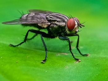
Cuterebra spp
Epidemiology: Arthropod dipteran parasite; some 34 different species in North America.
- Large maggots seen in the skin of dogs and cats and represent the larva of the rodent and rabbit bots. Adult flies lay eggs around the entrances to rodent burrows, and when host passes, the small first-stage maggot hatches and jumps onto the passing host.
- Larva capable of entering the host through the mouth, nose, eyes, or anus.
- In rodent hosts, larvae remain as 1st-stage larvae within the nasopharyngeal region near the posterior end of the soft palate and in the nasal passages. Maturation time from infection to when the larva leaves the warble is between 3 to 8 weeks.
- Seasonality: Adult flies have single emergence in late spring in the more temperate parts of the United States.
- Cats: Infected as they hunt. Kittens may be infected by larvae brought back on the fur of the queen.
- How larvae enter cats unknown, but probably through the mouth, nose, or anus as they do in the rodent and lagomorph hosts.
Clinical signs: Depend mainly on where the larva locates.
- Skin lesions; (warble) containing a single larvae usual present on the cheek, neck, or back, but other sites are reported.
- Acute upper respiratory tract distress; severe sneezing, unilateral initially serous then mucopurulent nasal discharge. Sneezing and nasal discharge can persist for a week.
- May be accompanied by unilateral facial swelling especially over the nose.
- Bloody nasal discharge, soft palate and pharyngeal swelling have been reported.
- Laryngeal edema (larval migration in the cervical neck) may cause laryngeal edema and arrest. Respiratory signs may persist for few days to several weeks, then often recede in severity occasionally to be followed by neurologic signs
- If neurologic signs develop, they will do so one to two weeks after respiratory signs, although respiratory signs have been reported to occur as long as 4 to 10 weeks before the onset of neurologic signs.
Diagnosis: Viewing the larvae within the respiratory tract, or made on circumstantial evidence of acute rhinitis (sometimes progressing to neurologic disease) in an outdoor cat during late summer and fall. The larvae or its migration tracts through the brain may be identified on CT scan or MRI.
Treatment: Ivermectin (0.1 to 0.3 mg/kg, PO, q24h, 3 days) very effective.
- Prednisone (1 mg/kg, PO, q12h, 3 weeks, then 1 mg/kg, PO, q24h, 3 weeks). [Diagnosis based on clinical signs, but usually improve). Few develop neurologic disease. Some neurologic cats improve clinically with this treatment but outcome unchanged.
Pneumonyssoides caninum (canine nasal mite)
Epidemiology: Arthropod parasite of nasal sinuses of dogs. Occurs in the USA, Canada, Japan, Australia, Sth. Africa and Europe.
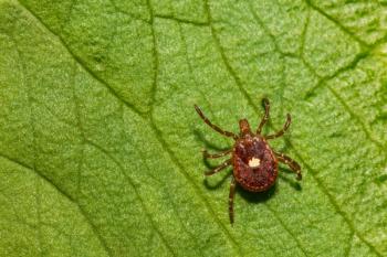
- Adult mites (1 mm X 0.5 mm in size) are easily identified by their leg morphology; the first pair of legs each terminate in a pair of large hooks, while legs two, three, and four each terminate in a sucker armed with a pair of smaller hooks.
- Believed that dog-to-dog transmission is by the direct transfer of larvae from one infested dog to another.
Clinical signs: Sneezing, although can present with facial pruritis, snuffling, snorting + nasal discharge and excessive lacrimation.
Treatment: Ivermectin (200 µm/kg body weight, PO or SC) or Milbemycin (0.5 mg/kg, PO) once weekly for 3 weeks are effective.
Linguatula serrata
Epidemiology: Pentastomid parasites that represent a group of specialized crustacean-like arthropods.
- Adult female is approximately 8 to 10 cm long and 1 cm in diameter; the male is about 2 cm long. The body appears superficially annulated, and the worms tend to be tan to brown in color.
- Life cycle requires an intermediate host. The eggs that are passed by the female contain a four-legged larvae. Eggs do not appear in the feces of the dog but instead are found in the nasal secretions
- Suspected that most dogs obtain their infections by the ingestion of sheep offal. When dogs ingest infected tissues, the nymphs migrate up the back of the throat into the nasal turbinates. Once swallowed, the nymphs do not migrate back up the esophagus.
- Prepatent period (PPP) is about 6 months. Adult worms live about 2 years.
Clinical signs: Sneezing, slight nasal discharge sometimes containing blood. The parasites become large, lie in the recesses of the nasal turbinates, and attach themselves firmly to the mucous membranes with their four hook. The adults apparently feed on respiratory mucosal cells and blood. When fully grown, the parasites are capable of causing nasal obstruction.
- Humans may also become infected but not from dogs.
Diagnosis: Eggs (yellowish oval, 80 m, surrounded by bladder-like envelope and containing a four-legged larva) in the nasal secretions. - identifying larvae during rhinoscopy.
Treatment: Physical removal only treatment described. Ivermectin (200 µg/kg, PO, once) may be efficacious.
Eucoleus boehmi
Epidemiology: Capillarid nematode parasite of nasal mucosa of the dog. Adult worms live threaded through the mucosa of the nasal sinuses. Adults appear as very fine threads seen grossly as very fine transparent hairs.
Clinical signs: Sneezing, nasal discharge.
Diagnosis: Identifying eggs in feces (eggs can be recovered in nasal washings).
Treatment: Fenbendazole (50 mg/kg, PO, q24h, 7 days). Ivermectin (200 µg/kg, PO, once).
Parasites of the lung parenchyma
Aelurostrongylus abstrusus
Epidemiology: Metastrongyloid nematode parasite of cats.
- Adult female (9 to 10 mm long) and males (4 to 6 mm) worms coil in the terminal respiratory bronchioles and alveolar ducts. The females lay eggs that contain a single cell when laid and which embryonate within the alveolar ducts and the surround alveoli. The larvae hatch from the eggs, are carried up the ciliary escalator, swallowed, and passed in the feces.
- Larvae: approx. 360-390 um long, characteristic dorsal spine on the tail.
- Cats infected when ingest infected snail intermediate host or, more likely, paratenic hosts (mice, birds).
Clinical signs: Heavy infestations (100 larvae) can cause severe pulmonary disease and radiographic changes by 2-wks PI. Most severe disease occurs 5 to 15 weeks after infection.
- Presents as alveolar lung disease (no pulmonary hypertension or associated RVH disease).
- Most infections are asymptomatic with the cat recovering uneventfully.
- May get signs of severe bronchopneumonia (rapid open-mouthed abdominal breathing with) - eosinophilia rare. - radiographs: diffuse interstitial pattern. After a week of treatment, the radiographic pattern may appear worse (more peribronchial infiltrates with areas of alveolar consolidation) in spite of clinical improvement in signs.
Diagnosis: Identifying typical larvae in the feces or in a trans-tracheal wash.
Treatment: Fenbendazole (20 mg/kg, PO, q24h, for 5 days, then repeat after 1 week).
- Ivermectin (400 µg/kg, PO, once, followed by a second dose of 400 µg/kg, PO, one week later).
- Prednisone (1 mg/kg, PO, q12h, 5 days) alleviates signs during recovery.
Eucoleus aerophilus (Capillaria aerophila)
Epidemiology: Trichuroidea parasite with bi-operculate eggs. Direct life cycle.
- Eggs – easily confused with those from other Capillarids (Eucoleus boehmi of the nose, and Pearsonema plica in the urinary bladder) and whipworms of dogs (Trichuris vulpis).
- Adult worms – embed in the mucosal lining of large airways expelling eggs into the respiratory passages. Eggs - coughed up the trachea, and swallowed to be passed in the feces.
- Infection – occurs by ingesting L1 larvae (take about 40 days to mature in eggs).Infections - can last as long as a year. PPP = 3 to 5 weeks.
Clinical Signs: Fairly common infection in both cats and dogs.
- Most infections are asymptomatic - rarely causes clinical signs.
- When signs occur:- mild wheezing, chronic cough can occur. Very rarely produces weight loss. When complicated with bacterial pneumonia, can cause death.
- Thoracic radiographs may show diffuse mild bronchoalveolar pattern but are not pathognomonic.
- Diagnosis made by finding bi-operculate eggs in feces or tracheal wash fluids.
Treatment: Assymptomatic cases do not require treatment.
- Fenbendazole (50 mg/kg, PO, q24h, 14 days). Treatment of choice in dogs.
- Ivermectin (200 µg/kg, PO, once). Efficacy is unknown but is effective against nasal capillariasis and indications are that it is effective against E. aerophilus as well.
Filaroides hirthi
Epidemiology: Metastrongyloid nematodes found in the lung parenchyma of dogs. Direct life cycle.
- Infection of pups probably occurs during nursing. After ingestion, larvae migrate to lungs via hepatic-portal or mesenteric lymph system. Prepatent period is 5 weeks. Larvae appear in the feces.
- Most cases reported in beagles in research colonies.
Clinical signs: Nonproductive cough + increased respiratory rate.
- Severe infestations: respiratory distress and exercise intolerance, looks like "kennel cough."
- Radiographs: diffuse interstitial lung opacities and mixed alveolar patterns with consolidation
Treatment: Albendazole (25 mg/kg, PO, q12h, 5 days, then repeat treatment 2 wks later).
- Fenbendazole (50 mg/kg, PO, q12h, 14 days), or Ivermectin (0.2 mg/kg, PO, q24h, 3 days).
- Prednisone (1.25 mg/kg, PO, q24h, 14 days)
Paragonimus kiettiella
Epidemiology: Trematode (fluke) normally found in mink, but occasionally in the lungs of dogs and cats.
- Adult pairs live in subpleural cysts that communicate with bronchiole. Eggs produced in the cysts are carried into the bronchiole, swept up airways and swallowed.
- Carnivores infected by eating 2nd intermediate host (crayfish).
Clinical signs: Most are asymptomatic but those with disease present with a chronic cough (unresponsive to most treatments) and rarely pneumothorax.
Diagnosis: Identifying eggs (large, operculated) in feces or tracheal wash. Radiology; multiloculated cysts (dogs), and interstitial nodules (cats).
Treatment: Praziquantel (23 mg/kg PO, q8h, 3 days), or fenbendazole (50 mg/kg PO, q24h, 10-14 days).
Trachea and bronchi
Crenosoma vulpis
Epidemiology: Metastrongyloid nematode of bronchi (dogs and other canids). Adult worms (males; 4 to 8 mm long, females; 12 to 16 mm long) parasitize the terminal bronchi of the respiratory tract where eggs are laid, develop and hatch, larvae then coughed up and swallowed to be passed in the feces.
- Dogs infected when ingest gastropod (snails) intermediate hosts. Larvae migrate to the lungs by way of the visceral lymphatic or via the hepatic portal system: prepatent period about 18 to 21 days.
Clinical signs: Infection occurs during summer in dogs living or visiting rural areas frequented by foxes, the more usual definitive host. - dry, nonproductive cough easily elicited by tracheal palpation. Cough may be chronic and productive.
- Radiology: Diffuse bronchial patterns with prominent interstitial markings.
- Bronchoscopy: Moderate mucoid to mucopurulent discharge in the airways. Cytology of tracheal wash: reveal inflammatory cells, mainly eosinophils (matching a peripheral eosinophilia).
Diagnosis: Larvae (pointed tail, 250 - 300 um) in feces or tracheal wash sample.
Treatment: Fenbendazole (50 mg/kg PO, q24h, 3 days, or 20 mg/kg, PO, q24h, 14 days).
- Levamisole (7.5 mg/kg, PO, once, followed by a second dose 2 days later).
Oslerus osleri
Epidemiology: Metastrongyloid nematode: causing nodules in the terminal trachea and bronchi of dogs and other canids (coyotes, foxes). Adult worms found in subepithelial fibrous nodules usually close to the bifurcation of the trachea and extending down into the mainstem bronchi. Thin-shelled eggs or larva (L1 - infective stage and very similar to F. hirthi) are coughed up, swallowed, enter feces, or coat regurgitated food.
- Direct lifecycle. Pups infected by L1 in sputum (infected when cleaning, or coating regurgitated food.
Clinical signs: Dry cough (often precipitated by exercise or tracheal palpation) unresponsive to antibiotics or
steroids.
- Severe respiratory distress due to upper airway obstructive disease.
Diagnosis: L1 in fecal samples or in transtracheal washes. Bronchoscopy: identifies the brown reddy nodules at the tracheal bifurcation. Radiology may reveal soft tissue nodular densities at the tracheal bifurcation.
Treatment: Physical removal of nodules via bronchoscopy can be life saving.
- Oxfendazole (10 mg/kg PO, q24h, 28 days). Only drug known to work.
- Prednisone (0.5 - 1 mg/kg PO, q12h, 5 days post-nodule removal). Removes inflammation with worm death.
References
Glass EN, Cornetta AM, deLahunta A, et al. Clinical and clinicopathologic features in 11 cats with cuterebra larvae myiasis of the central nervous system. J Vet Intern Med 1998;12:365-368.
Gunnarsson LK, Moller LC, Einarsson AM, et al. Clinical efficacy of milbemycin oxime in the treatment of nasal mite infection in dogs. J Am Anim Hosp Assoc 1999;35:81-84.
Barr SC. Feline Lungworm (Aelurostrongylus). In: Canine and Feline Infectious Diseases and Parasitology. Barr SC and Bowman DD (eds). Ames Ia: Blackwell Publishing. 2006. p. 233.
Peterson EN, Barr SC, Gould WJ, et al. Use of fenbendazole for treatment of Crenosoma vulpis in a dog. J Am Vet Med Assoc 1993;202:1483-1484.
Kelly PJ, Mason RP. Successful treatment of Filaroides osleri infection with oxfendazole. Vet Rec 1985;116:445-446.
Barr SC, Lavelle RB, Harrigan KE, et al. Oslerus (Filaroides) osleri in a dog. Aust Vet J 1986;63:334-336.
Newsletter
From exam room tips to practice management insights, get trusted veterinary news delivered straight to your inbox—subscribe to dvm360.


