Risk factors and monitoring to avoid acute renal failure (Proceedings)
Acute kidney injury often results from ischemic or toxic insults and usually affects the most metabolically active tubular portions of the nephron.
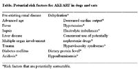
Acute kidney injury (AKI) often results from ischemic or toxic insults and usually affects the most metabolically active tubular portions of the nephron. If the ischemic or toxic insult is severe enough, acute renal failure (ARF) may result. In many cases, AKI and ARF inadvertently develop in the hospital setting in conjunction with diagnostic or therapeutic procedures. For example, renal damage may result from decreased renal perfusion associated with anesthesia and surgery or with the use of nonsteroidal anti-inflammatory drugs (NSAIDs). Similarly, renal damage may occur in patients treated with potential nephrotoxicants like gentamicin, amphotericin, and cisplatin. The nephron damage that occurs with ischemic or toxic insults is not always reversible; animals that do recover adequate renal function usually require prolonged and expensive intensive care. Several retrospective studies have documented the poor prognosis associated with ARF in dogs and cats. In a study of hospital-acquired ARF, the survival rate was only 40%. In another retrospective study of 99 dogs with all types ARF, 22% died, 34% were euthanized, 24% survived but progressed to chronic kidney disease (CKD), and only 19% regained normal or adequate renal function. Similarly, in a retrospective study of 32 cats with all types of ARF, 16% died, 31% were euthanized, 28% survived but progressed to CKD, and only 25% regained normal or adequate renal function. These studies underscore the importance of early detection of AKI and prevention of ARF. Several risk factors have been identified that predispose dogs to gentamicin-induced ARF (Table), however it is likely that many of these risk factors also predispose dogs and cats to other types of toxicant-induced ARF as well as ARF induced by ischemia. A combination of decreased renal perfusion and/or use of nephrotoxic therapeutic agents superimposed on more chronic, pre-existing risk factors is usually responsible for AKI/ARF in the clinical setting. Early detection of AKI facilitates appropriate intervention that can arrest or at least attenuate tubular cell damage and the development of established ARF.

Table. Potential risk factors for AKI/ARF in dogs and cats
Pathophysiology
Acute renal failure has three phases, which are categorized: 1) initiation, 2) maintenance, and 3) recovery. The initial insult occurs resulting in sub-lethal cellular injury in the initiation phase. Therapeutic measures started during this initiation phase may reduce the renal insult and prevent development of established ARF. The maintenance phase is characterized by tubular cell death and established nephron dysfunction. Therapeutic intervention during the maintenance phase, although often life saving, usually does little to diminish existing renal lesions or improve renal function. The recovery phase is the period when renal lesions resolve and renal function improves. Tubular damage may be reversible if the tubular basement membrane is intact and viable epithelial cells are present. Although additional nephrons cannot be produced and irreversibly damaged nephrons can not be repaired, functional hypertrophy of surviving nephrons may adequately compensate for the decrease in nephron numbers. Even if renal functional recovery is incomplete, adequate function may be reestablished in some cases.
Risk factors for AKI/ARF
Dehydration and volume depletion are perhaps the most common and most important risk factors for ARF (Table). Studies in human beings indicate volume depletion increases a patient's risk of developing ARF by a factor of ten. Hypovolemia not only decreases renal perfusion, but also decreases the volume of distribution of nephrotoxic drugs and results in decreased tubular fluid flow rates and enhanced tubular absorption of toxicants. In addition to hypovolemia, renal hypoperfusion may be caused by decreased cardiac output, decreased plasma oncotic pressure, increased blood viscosity, systemic hypotension, and decreased renal prostaglandin synthesis. Any of these conditions can increase the risk of ARF in the hospital setting.
Pre-existing renal disease and advanced age, which is often associated with some degree of decreased renal function, can increase the potential for nephrotoxicity by several mechanisms. The pharmacokinetics of potentially nephrotoxic drugs can be altered in the face of decreased renal function. Gentamicin clearance has been shown to be decreased in dogs with sub-clinical renal dysfunction, and the same is probably true for other nephrotoxicants. Animals with renal insufficiency also have reduced urine concentrating ability and, therefore, decreased ability to compensate for prerenal influences. Renal disease may also compromise production of prostaglandins that help maintain renal vasodilation and blood flow.
Decreased serum concentrations of several electrolytes can increase the risk of ARF. For example, hyponatremia potentiates IV contrast media-induced ARF in dogs. Additional studies in dogs have demonstrated that dietary potassium restriction exacerbates gentamicin nephrotoxicity, possibly because potassium depleted cells are more susceptible to damage. It is important to note that gentamicin administration in dogs is associated with increased urinary excretion of potassium. This increased urinary excretion of potassium could result in reduced intracellular concentrations and increased nephrotoxicity in clinical patients. Therefore, serum electrolyte concentrations should be closely monitored in patients receiving potentially nephrotoxic drugs, especially if these patients are anorexic, vomiting, or have diarrhea.
Administration of potentially nephrotoxic drugs or drugs that may enhance nephrotoxicity obviously increases the risk of ARF. For example, concurrent use of furosemide and gentamicin in dogs is associated with increased risk and severity of ARF. Furosemide probably potentiates gentamicin-induced nephrotoxicity by causing dehydration, reducing the volume of distribution of gentamicin, and increasing the renal tubular absorption of gentamicin. Fluid therapy minimizes, but does not avoid, the potentiating effect of furosemide on gentamicin nephrotoxicity in the dog, because furosemide also facilitates cellular uptake of gentamicin independent of hemodynamic changes. By similar mechanisms, furosemide has been shown to enhance intravenous radiocontrast-induced nephrotoxicity in human beings. Use of NSAIDs can also increase the risk of ARF. Anesthesia, sodium and/or volume depletion, sepsis, congestive heart failure, nephrotic syndrome, and hepatic disease are conditions in which prostaglandin-induced renal vasodilatation becomes important and the susceptibility to NSAIDs is increased.
Evidence in dogs suggests that the quantity of protein fed prior to a nephrotoxic insult can significantly affect the subsequent renal damage and dysfunction. High dietary protein prior to and during gentamicin administration reduces nephrotoxicity, enhances gentamicin clearance, and results in a larger volume of distribution compared with medium or low dietary protein. The beneficial effects of high dietary protein are likely associated with increased glomerular filtration and, therefore, improved toxicant excretion. High dietary protein also results in increased urinary excretion of protein, which may compete for nephrotoxicant reabsorption by tubular epithelial cells. Increasing dietary protein ahead of time may not be a viable clinical option but dogs with reduced protein intake should be recognized as having increased risk for developing ARF. Once renal damage has occurred, high dietary protein would likely result in increased serum urea nitrogen and phosphorus concentrations and, therefore, would not be recommended.
Risk factors are additive and any complication occurring in high risk patients increases the potential for ARF. Patients with shock, acidosis, sepsis, and major organ system failure are at increased risk for ARF, and these are the patients that are likely to require aggressive treatment including prolonged anesthesia, surgery, or chemotherapeutics which are potentially damaging to the kidneys. As an example, ARF is relatively common in dogs with pyometra and E. coli endotoxin-induced urine concentrating defects, especially if fluid therapy is inadequate during anesthesia for ovariohysterectomy or during the recovery period. Trauma, extensive burns, vasculitis, pancreatitis, fever, diabetes mellitus, and multiple myeloma are additional conditions associated with a high incidence of ARF in veterinary medicine.
Early recognition of kidney injury/dysfunction
Since therapeutic intervention is most successful when initiated during the induction phase of ARF, early recognition of renal damage/dysfunction is important. Physical examination of the patient at risk for ARF should include evaluation of cardiac rate, rhythm, pulse quality and hydration status. Monitoring body weight, packed cell volume and plasma total solids in comparison to baseline values may indicate subtle changes in hydration status during hospitalization. Blood pressure measurement will help identify hypotensive and hypertensive patients, both of whom may be at increased risk for renal injury.
Numerous urine parameters can herald the development of ARF. Urine output should be monitored in all high-risk patients that undergo anesthesia. Urine production may be quantitated using a closed indwelling catheter collection system. Normal urine output is approximately 1-2 ml/hr/kg body weight. Oliguria (< 0.25 ml/hr/kg) or anuria requires prompt attention and treatment. Nonoliguric ARF is being recognized with increasing frequency; increases in urine production, therefore, may also signal the onset of renal damage. Examples of non-oliguric ARF include those induced by gentamicin and cisplatin. In animals receiving potentially nephrotoxic drugs, increased urine turbidity or changes in urine sediment (increasing numbers of WBCs, RBCs, renal epithelial cells, or cellular or granular casts) may be indications of acute renal tubular damage. Finally, the acute onset of normogylcemic glucosuria, proteinuria, or increases in the fractional clearance of sodium and chloride may also be indicative of early tubular damage. The interpretation of all of the above parameters is enhanced by knowledge of baseline values.
Detection of enzymes such as gamma-glutamyl transpeptidase (GGT) and N-acetyl-beta-D-glucosaminidase (NAG) in the urine has proven to be a sensitive indicator of renal tubular damage. These enzymes are too large to be normally filtered by the glomerulus, and, therefore, enzymuria indicates cell leakage, usually caused by tubular epithelial damage or necrosis. Urinary GGT originates from the proximal tubule brush border and NAG is present in proximal tubule lysosomes. In a study of gentamicin-treated dogs, increased urinary GGT activity was the earliest marker of renal damage/dysfunction. Interpretation of enzymuria is aided by comparison to baseline values obtained prior to a potential renal insult; 2 to 3-fold increases over baseline suggest significant tubular damage. Urine enzyme/creatinine ratios from spot urine samples have been shown to be accurate in dogs prior to the onset of azotemia obviating the need to for timed urine collections. False positive results can occur with severe glomerular damage, resulting in increased glomerular filtration of serum enzymes. False negative results can occur after tubular damage depletes tubular enzyme stores. In those cases where treatment with an aminoglycoside is thought to be necessary, measurement of serum antibiotic trough concentrations may help predict the onset of ARD. Increasing serum trough aminoglycoside concentrations are associated with increased risk of nephrotoxicity. If trough concentrations increase above 2 µg/dl for gentamicin or above 5 µg/dl for amikacin, the antibiotic should be discontinued or the time between doses should be increased. Administering the same total daily dose once a day vs. dividing the daily dose and administering them multiple times per day will typically result in lower serum trough concentrations and reduced nephrotoxicity.
Summary
Knowledge of the predisposing risk factors allows the clinician to assess the risk-benefit ratio in individual cases in which an elective anesthetic procedure is considered or the use of potentially nephrotoxic drugs is indicated. In some cases, predisposing risk factors can be corrected prior to any potential renal insults. In other cases such as geriatric patients with suspected pre-existing renal disease, more intensive monitoring of the patient may allow detection of AKI/ARF in its early phase prior to the onset of established failure.