Surgery STAT: Scrotal ablation for routine and cryptorchid castration in horses
A collaborative column between the American College of Veterinary Surgeons and DVM Newsmagazine.
EDITOR'S NOTE: SurgerySTAT is a collaborative column between the American College of Veterinary Surgeons (ACVS) and DVM Newsmagazine.
This month, Charles T. McCauley writes about scrotal ablation for routine and cryptorchid castration in horses. In January, Karen M. Tobias, DVM, MS, Dipl. ACVS, professor of small animal surgery at the University of Tennessee, discusses diagnosis and care of methicillin-resistant wound infections (MRSP).
To locate a diplomate, ACVS has an on-line directory, which includes practice setting, species emphasis and research interests (www.acvs.org/VeterinaryProfessionals/FindaSurgeon ).
Historically, cryptorchid castration has been performed utilizing two incisions, one near or directly over the inguinal ring of the retained testicle and the second adjacent to the median raphe for removal of the normally descended testicle. The inguinal incision is closed primarily or packed with roll gauze, which requires the gauze or the suture to be removed at a later date.

Dr. McCauley
The following describes a technique for scrotal ablation in routine and cryptorchid castrations utilizing a single incision that is closed without skin suture or staples. I use this technique frequently when performing routine castration on horses. Using this technique, I see fewer postoperative complications and an earlier return to training in comparison with routine castration technique.
Surgical technique
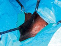
With the horse anesthetized, the prepuce is packed with gauze and closed with continuous suture or towel clamps. The skin surrounding and directly over the scrotum is prepared for aseptic surgery. The scrotum is grasped on the median raphe with two Allis tissue forceps positioned 6–8 cm apart (Photo 1).
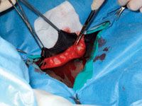
Using the forceps, the skin is elevated and an 8–10 cm fusiform incision is made at the margin of the elevated skin. The scrotal skin is placed under tension, and the scrotal fascia and tunica dartos are separated from the underlying testicle and surrounding skin using a combination of blunt and sharp dissection. These tissues are then removed en bloc (Photo 2).

Photo 1: Elevation of the scrotal skin with Allis tissue forceps.
If minor hemorrhage occurs, it is controlled by temporary application of hemostats. The skin surrounding the incision is loose and highly mobile, allowing the incision to be manipulated over the right or left inguinal ring to access a retained testicle. The cryptorchid testicle is located and removed using the previously described technique. Thereafter, the skin incision is manipulated over the descended testicle, which is exposed and removed.
In my practice, both the cryptorchid and normally descended testicle are removed by a routine closed castration with two minor variations. One variation is separation of the cremaster muscle away from the spermatic cord and emasculation of the muscle.
This allows a greater length of the spermatic cord to be exteriorized from the inguinal ring by eliminating the pull of the muscle on the cord. The second variation is placement of a single transfixation ligature using size 1 polydioxanone suture at the most proximal aspect of the spermatic cord, which is then emasculated distal to the ligature.
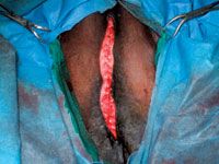
After the testicles have been removed, the deep fascia on either side of the incision and the remaining median raphe are closed using size 0 polyglactin 910 in a simple continuous pattern (Photo 3).
The skin edges are apposed using size 2-0 polyglactin 910 in a subcuticular pattern. Tissue adhesive often is applied as an added barrier to contamination; however, no skin sutures or staples are needed.
Discussion
Although the technique is slightly different than that previously described, I believe the proposed advantages of this technique and my results are similar to those previously reported by Palmer and Passmore.

Photo 2: Dissection for en bloc removal of the scrotal skin, scrotal fascia and tunica dartos.
Due to the high mobility of the scrotal skin, both inguinal rings and the descended testicle can be accessed through a single incision. When closed, there is minimal to no tension on the apposed skin edges; therefore, primary skin closure is not necessary. There is no postoperative drainage, and, subjectively, there is a significant reduction in the amount of postoperative preputial edema compared to routine castration techniques.

Photo 3: Completed closure of the subcutaneous tissue.
This technique minimizes the risk of post-castration eventration. Complete removal of the spermatic cord at the level of the inguinal ring and ligation of the spermatic cord reduce the risk of scirrhous cord formation.
In my hands, horses castrated by this technique typically return to full work within five to 10 days. To date I have had no complications using this technique. The primary disadvantages when using this procedure for routine castration include a longer surgical time and a modest increase in the cost of the procedure.
Dr. McCauley is an ACVS board-certified surgeon and an assistant professor of surgery in the School of Veterinary Medicine, Louisiana State University, Baton Rouge, La.
Visit dvm360.com/surgeryto access articles on veterinary surgery.
Suggested Reading
• Rodgerson DH, Hanson RR. Cryptorchidism in horses. Part II. Treatment. Compend Contin Educ Pract Vet. 1997;19(12):1372-79.
• Adams SB, Fessler JF. Castration. In: Atlas of Equine Surgery. Philadelphia PA: WB Saunders Co, 2000:209-214.
• Palmer SE, Passmore JL. Midline scrotal ablation technique for unilateral cryptorchid castration in horses. J Am Vet Med Assoc. 1987;190(3):283-5.


