The tetraparetic dog: The upper motor neuron, the lower motor neuron, and the in-between (Proceedings)
Tetraparesis or tetraplegia is a neurological condition in which all four limbs are weak (paresis) or paralyzed (plegia).
Tetraparesis
Tetraparesis or tetraplegia is a neurological condition in which all four limbs are weak (paresis) or paralyzed (plegia). A very long list of differential diagnoses could be made in the case of tetraparesis/plegia; however, the results of the neurological examination, along with history and signalment make defining differential diagnoses much more simple. For example, the neurological examination for an animal with a condition of generalized lower motor neuron disease (such as tick paralysis, botulism and polyradiculoneuritis) will be very different from an animal that has a cervical (C1-C5) or cervicothoracic (C6-T2) syndrome. Defining the lesion localization allows a more defined list of diseases. The most common localizations for a tetraparetic/plegic animal are: C1-5 spinal cord, C6-T2 spinal cord, and generalized lower motor neuron disease.
The definition of the lower motor neuron is the nerve cell body (soma), the peripheral axon and the nerve terminal that directly innervates the skeletal muscle. These neurons, also called α-motoneurons are found throughout the spinal cord in the ventral horn gray matter from C1-Cy (coccygeal) levels. Some of the brain stem nuclei can also be considered lower motor neurons as they also directly innervate muscle (e.g. facial nerve, mandibular branch of the trigeminal nerve). The upper motor neuron is a nerve whose cell body originates in the cerebrum (corticospinal tracts) or brain stem (rubrospinal, reticulospinal and vestibulospinal tracts) and whose axon travels in lateral or ventral white matter funiculi. These axons ultimately terminate on α-motoneurons or the interneurons that interact with them to initiate voluntary movement. Some of the white matter, therefore, is the axons of these spinal motor systems that are the source of voluntary movement. Without information from the upper motor neuron, the lower motor neuron does not function to initiate movement on its own, except in very rare cases (spinal walking).
When discussing "upper motor neuron signs" and "lower motor neuron signs", we are referring to those signs that are seen in the limbs during neurological examination. While there are lower motor neurons throughout the spinal cord, we generally only test their function (the reflex arc) in the limbs. The lower motor neurons that innervate the thoracic limbs are found in the cervical intumescences (C6-T2) and those that innervate the pelvic limbs are found in the lumbosacral spinal cord (L4-S1/2). For a normal myotatic reflex, the reflex arc requires the peripheral nerve and spinal cord in this region to be intact. Injury to the peripheral sensory nerve, the spinal cord segments and/or peripheral motor nerve typically results in a decreased to absent reflex. Animals with spinal injury or peripheral nerve injury will often be paretic or plegic and have some degree of ataxia. These findings are not necessarily localized as they can occur from lesions in the intumescence or anywhere above it. Thus, it is the findings in the limbs on reflex testing and muscle tone that give us some idea of the lesion localization.

Clinical findings
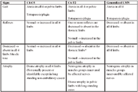
In the tetraparetic dog, the lesion localization will be differentiated by these findings. The C1-5 lesion can be separated from the C6-T2 lesion and both can be separated from the generalized lower motor neuron lesion.
C1-C5 Lesion Localization
In this region of the spinal cord, the lower motor neurons supply muscles of the neck. At this site the motor information that is affected are the white matter tracts (axons) of the upper motor neurons.
C6-T2 Lesion Localization
In the C6-T2, there are lower motor neurons that supply the thoracic limbs directly and upper motor neuron axons to the pelvic limbs traverse this region.
Generalized Lower Motor Neuron Localization
When there is injury or disease to a peripheral nerve, then the reflex arcs become affected and the reflexes are depressed to absent along with decreased strength and potentially sensory if both types of fibers are involved.
Less common localizations include generalized muscle disease, brain stem lesions and diffuse cerebral disease (such as metabolic diseases or increased intracranial pressure). Generalized muscle diseases are relatively uncommon and are not commonly acute in onset. They will typically be associated with stiff-stilted gaits, muscle atrophy and variable presence of muscle pain. Brain stem and cerebral diseases that are associated with tetraparesis with UMN signs and would obviously be associated with other signs associated with brain impairment such as abnormal mentation, seizures and cranial nerve signs.

Clinical signs and localizations
*With a C6-7 focal lesion, muscle tone may actually be increased in the thoracic limbs due to preferential disruption lower motor neurons of flexor muscles. Flexor withdrawal and/or biceps brachii reflexes will be decreased to absent.
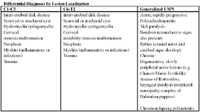
Once determined, the lesion localization, along with signalment and history will allow the development of a rational list of differential diagnoses. An appropriate differential diagnoses list will allow appropriate diagnostic testing, management and therapy.

Differential diagnoses by lesion localization