“We did a PET scan on horse with hock arthritis. The lameness improved after intra-articular anesthesia of the distal tarsal joints. The radiographs were unimpressive, but with PET, the subchondral bone in the distal tarsal joints ‘lit up’ beautifully,” said Garrett.
“With all advanced imaging modalities, we have a better way to find problems earlier,” emphasized Garrett. “A PET scan can pick up areas of abnormal bone metabolism much sooner than a radiographic image can. We can also see improvement in these areas over time with follow-up scans,” she added. “It is one more tool in our arsenal to find injuries sooner, treat them better, and hopefully prevent longer term potentially permanent damage from occurring,” stated Garrett emphatically.
PET scanners for racetrack horses
According to Jo O’Brion, RVT, Southern California Equine Foundation (SCEF) nuclear medicine technician, SCEF is the first racetrack hospital to have acquired equine PET pioneered at UC Davis. Located in Santa Anita Park, SCEF began using it in December 2019. This was shortly after a cluster of fatalities occurred at Santa Anita in 2019.1
Since then, SCEF came to realize the advantages PET would offer the racing industry, and to racetrack veterinarians worldwide.
“For a high interest venue, such as the Breeders’ Cup, with roughly 200 horses on the grounds before the event, we could PET scan every horse within a few days,” states Joseph Dowd, DVM, PhD, president of SCEF. "With an extra scanner, more staff, we could double our PET capacity, thereby allowing every single horse to be scanned prior to racing. Potentially, this would allow us not to have those catastrophic injuries during the Breeder’s Cup.
Breakthrough technology to visualize the lesion
During the summer of 2018, Spriet visited Del Mar, California to deliver a talk on early PET data obtained in anesthetized horses. “It was the first time we could see with imaging in live horses the small lesions of the sesamoid bone, that we’d known for [more than] 20 years were associated with breakdowns from the necropsy work of Dr. Sue Stover, DVM, DACVS, UCDavis, in conjunction with the CHRC (CA Horse Racing Board), who began cataloging fatal injuries [in] 1990,” stated Dowd. “Dr. Stover started to recognize a characteristic small lesion in the sesamoid bone and at the palmar aspect of the condyle bone, a site of bone remodeling, as being a potential precursor to fetlock breakdown, where overload arthrosis and subchondral bone failure occurs.”
“Without PET at that time, practitioners had no way of seeing/imaging that lesion for us to stave off catastrophic injury. Once Dr. Spriet perfected standing PET, we then had the technology to help us visualize these lesions.”
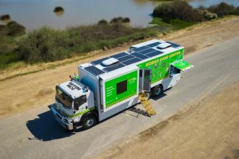
“PET is fast, we could do up to 20 horses per day. We can let a horse train in the morning, then come over to do a PET scan within a few minutes; put him in decontamination from the radioisotope and he can home back to the barn within a few hours. Therefore, you really don’t upset a horse’s life much at all.”
“As we began to do PET scans on racehorses, we realized PET would be better than other imaging modalities we’d used in racehorses," notes Dowd.
Additonally, O’Brion said, that funded by the Grayson Jockey Club Research Foundation, in conjunction with SCEF and Dolly Green Research Foundation (DGRC), SCEF has been able to complete various studies with its numerous populations of racehorses. “[This includes] comparison studies between PET and MRI and PET and scintigraphy; a study assessing the evolution of injuries in horses in lay-up or in training; a study on performing racehorses, to compare horses with visible clinical issues to horses that are performing well with no clinical symptomology; and currently a study of condylar fracture repair, following those horses through 90 days of healing post-surgery.”
“PET scans on racehorses have been invaluable,” added O’Brion. “We can do multiple PET scans per day. It takes on average 10 minutes to do, so horses can maintain a regular training schedule.”
“In addition to the equine PET scan, the SCEF hospital at Santa Anita also maintains a Gamma Camera and a standing MRI so that we can increase our through-put of diagnostics using multiple modalities at the same time. It increases our ability to get more horses scanned and potentially diagnosed.”
Along with the PET scanner at Santa Anita, similar scanners are available at UC Davis (also vanned to Golden Gate Fields); New Bolton Center (University of Pennsylvania School of Veterinary Medicine); Rood & Riddle Equine Hospital; and the World Equestrian Center (Ocala, Florida). Several more racing venues will likely install PET in the future.
“One of the advantages of PET in comparison with scintigraphy is [the ability] to better localize clinical pathology,” noted O’Brion. “Not only do some lesions show up earlier on PET than with scintigraphy, but it is also easier to quantify the ‘danger’ aspects of a lesion. With PET we’re able to look at specific areas of the condyle or sesamoid to note that is it either okay, or in danger of pathologic fracture.”
“As we’ve done repeated PET scans on the same horses, we’ve been able to gather sufficient data to enable our veterinarians to better tell trainers, that their horse needs some time off, and/or predict whether a lesion is likely to come back, or not.”
Takeaways
PET imaging technology is the newest equine imaging technology and detects small lesions that could not be seen with other imaging technologies. While PET is invaluable in the racetrack setting, as we gain more knowledge on the technology it is emerging as an advanced diagnostics tool for the entire equine sport industry. The goal is to perform PET scans of the fetlock in racehorses, and then determine from those images which horses are at higher risk. PET scans help ensure proper training schedules and horses succeed throughout their racing career.
Reference
Kane E. Racing surface at Santa Anita likely contributed to 25 equine deaths. March 17, 2019. Accessed July 28, 2022. https://www.dvm360.com/view/racing-surface-santa-anita-likely-contributed-25-equine-deaths


