Thymoma in an 11-year-old dog: Radiology perspective
Dr. Adrien Hespel provides the radiology perspective on this thymoma case.
The normal thymus is visualized in the cranioventral mediastinum in young dogs as an inverted wedge shape known as a “sail sign” (Figure 1). It is usually inconspicuous by 1 year of age because of thymic involution.1
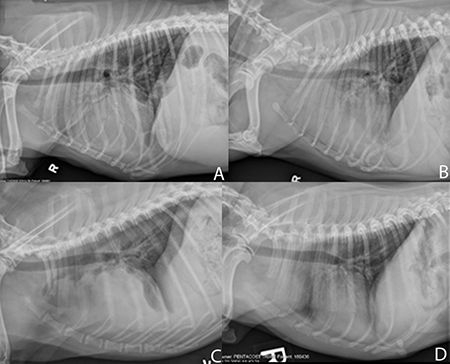
Figure 1. Four dogs with four different cranial mediastinal masses (none are the patient in this case): neuroendocrine tumor (A), lymphoma (B), hemangiosarcoma (C), thymoma (D). Note how similar all the masses are.In dogs, the cranial mediastinum should measure about twice the width of a vertebral body on a VD or DV projection. However, brachycephalic breeds can have a cranial mediastinum measuring less than twice the width of the vertebral column.1 The presence of a cranial mediastinal mass is unspecific for thymoma and can represent a wide range of processes both benign and malignant, including mediastinal cysts, abscesses, hematomas, granulomas, lymphomas, hemangiosarcomas, neuroendocrine tumors and mast cell tumors.

Dr. Adrien HespelThymomas may grow extremely large, and when they do, they tend to extend on the left of midline because of the orientation of the ventral recess of the cranial mediastinum.1,2 Because of their size, thymomas are usually associated with a significant mass effect, like any cranial mediastinal mass. On a lateral thoracic radiograph, the trachea may be displaced dorsally, and there may be caudal displacement of the cardiac silhouette and tracheal bifurcation. On a VD or DV radiograph, the trachea may be displaced to the right, and there may be caudal displacement of the lung lobes and widening of the cranial mediastinum.
Radiographically, in up to 40% of the cases of dogs with thymomas, a megaesophagus is concurrently seen.3 That is secondary to myasthenia gravis appearing as a paraneoplastic syndrome. In such cases, it is important to be vigilant for any signs of aspiration pneumonia.
Transthoracic ultrasonographic examination is a commonly performed procedure for further evaluation of a cranial mediastinal mass (Figure 2). Thymomas are commonly visualized as a heterogeneous irregular cystic mass with numerous cavitations. In this regard, thymomas differ from lymphomas, which tend to be more homogeneous and less cystic in nature.
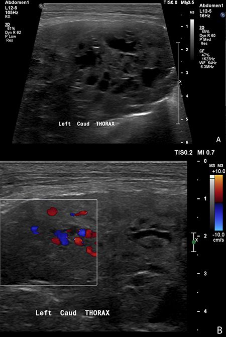
Figure 2. A B-mode ultrasonographic image (A) and a color Doppler ultrasonographic image (B) of a thymoma (not the patient in this case). The mass is cystic, complex and highly vascular.Advanced imaging such as computed tomography (CT) or magnetic resonance imaging (MRI) has been described for additional evaluations of thymomas (Figures 3A-3C). The primary purpose of these examinations is to help with surgical planning and to further evaluate for metastatic disease. In one retrospective study of dogs with thymomas, 15% had evidence of locoregional lymph node enlargement, 15% had evidence of neoplastic vascular invasion in the cranial vena cava, and 5% had evidence of pulmonary metastasis.3 It should also be noted that about 27% of dogs with thymoma had an additional nonthymic tumor.
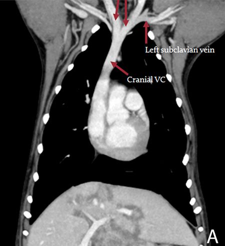
Figure 3A. Dorsal multiplanar reconstruction of a post-contrast CT of a normal dog.
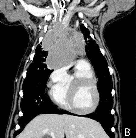
Figure 3B. Dorsal multiplanar reconstruction of a post-contrast CT of a dog with a large thymoma (not the patient in this case). Note the heterogeneity of the mass and the invasion in the vessels cranially contiguous to the mass.
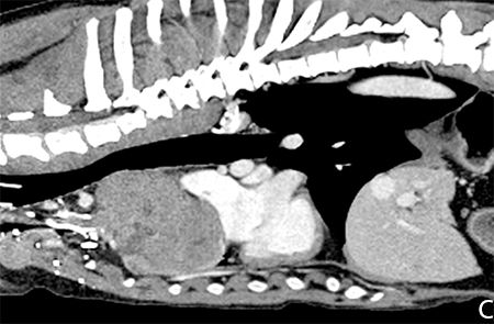
Figure 3C. Sagittal multiplanar reconstruction of a post-contrast CT of a dog with a large thymoma (same as Figure 3B). The vascular invasion is again noted. The mass deviates the trachea dorsally and is intimately associated with the cranial aspect of the thorax.On CT, thymomas can have a cystic center with an irregularly marginated, thick, irregular internal wall. When it comes to evaluating cranial mediastinal masses, it has been demonstrated that nonangiographic CT significantly underestimates the presence of vascular invasion compared with surgery.4 Thus, if CT is to be performed, ideally, an angiographic study would also be obtained. CT-guided tissue sampling can also be obtained.
Only one study reported the use of MRI for the evaluation of canine thymoma in a total of five dogs.5 In those cases, the masses were reported as T1 and T2 hyperintense when compared with muscles, with foci of T2 hyperintensity being T1 isointense to hypointense. After contrast administration (gadolinium), diffuse heterogeneous contrast enhancement of the mass with small nonenhancing foci was noted.
References
1. Thrall DE. Textbook of veterinary diagnostic radiology. 6th ed. St. Louis: Elsevier Health Sciences, 2013.
2. Wisner E, Zwingenberger A. Atlas of small animal CT and MRI. Ames, Iowa: John Wiley & Sons, 2015.
3. Robat CS, Cesario L, Gaeta R, et al. Clinical features, treatment options, and outcome in dogs with thymoma: 116 cases (1999-2010). J Am Vet Med Assoc 2013;243(10):1448-1454.
4. Yoon J, Feeney DA, Cronk DE, et al. Computed tomographic evaluation of canine and feline mediastinal masses in 14 patients. Vet Radiol Ultrasound 2004;45(6):542-546.
5. Adams WH, Hecht S, Conklin GA, et al. MRI and MRA characterization and preoperative evaluation of thymomas in dogs. Vet Radiol Ultrasound 2013;54(4):408.
Podcast CE: A Surgeon’s Perspective on Current Trends for the Management of Osteoarthritis, Part 1
May 17th 2024David L. Dycus, DVM, MS, CCRP, DACVS joins Adam Christman, DVM, MBA, to discuss a proactive approach to the diagnosis of osteoarthritis and the best tools for general practice.
Listen