Ticks and tick-borne diseases (Proceedings)
Tick borne diseases are extremely important and emerging diseases in the United States.
Tick-borne diseases are extremely important and emerging diseases in the United States. The area in which you live will influence the diseases that are circulating in the environment. Although diseases such as Lyme disease has received a great deal of attention, other important diseases including ehrlichiosis, Rocky Mountain spotted fever, anaplasmosis and cytauxzoonosis have been emerging in various areas. A good travel history is imperative given various species of ticks and tick-borne diseases are more common in certain geographical areas. More information on tick-borne disease distribution can be found at
Diagnosis of tick-borne diseases: serology vs PCR
Testing is warranted on animals with the aforementioned clinical signs. PCR testing (detection of pathogen DNA) is more sensitive than serology for detection of Rocky Mountain spotted fever and Anaplasma/Ehrlichia species during the acute phase of the disease, prior to the development of an antibody response. Therefore, if an animal presents with acute signs suggestive of tick-borne disease (i.e. fever, lethargy, thrombocytopenia, leukopenia, arthropathy, neurologic dysfunction), the best test for diagnosis is the PCR. Whole blood (EDTA) should be obtained for the test, prior to antibiotic administration.
Serology is useful for detection of chronic/persistent infections, during which the numbers of pathogens are lower or absent from circulation and cannot be detected as easily by PCR. This is particularly true for Lyme disease. This organism localizes in the tissues and is difficult to detect in the blood. It is important to note that antibodies to these tick-borne agents may persist for several months to years (especially for E. canis), so detection of antibodies does not distinguish current infection from previous exposure. Also, high seroprevalence to these agents has been documented in healthy dogs in endemic areas, such as the Southern USA, and most dogs exposed to Anaplasma or Ehrlichia species will not develop overt clinical disease. Therefore, PCR of skin or other tissues (not blood) and/or complete blood count is useful to determine if seropositive animals are currently infected and have clinical disease.
Identification of ticks
Tick bodies are divided into two primary sections including fused head and thorax and abdomen. All adult and nymphal forms have 4 pairs legs and no antennae and all larval forms have 3 pairs of legs. The importance of determining larvae vs other stages include to determine the likelihood of tick being infected with various pathogens. Unless transovarial transmission occurs, larvae are unlikely to be infected with pathogens, while nymphs and adults have higher likelihood include with pathogens in transstadial transmission. Whereas hard ticks have scutum, soft ticks do not have scutum. Ticks are great vectors due to their ability to be persistent blood-suckers which attach firmly & feed slowly, long life spans, may be geographically widespread, resistant to environmental conditions, high reproductive potential, and can pass infective agents through egg to next generation and/or through successive stages. Ticks bites in themselves can lead to wounds and Inflammation from salivary proteins. Secondary infection and disease can be due to toxicosis, local necrosis, and tick paralysis. Tick bites predispose animals to secondary attacks by myiasis-producing flies.
Soft tick have no scutum are soft, tough, leathery body, do not stay attached-instead take multiple small volumes of blood, and often feed at night.
Soft ticks include Otobius megnini (Spinose Ear Tick) transmits relapsing fever caused by a Borrelia spp. (different than Borrelia burgdorferi which causes Lyme Disease). Spinose ear ticks are more common in western states that are west of 100th meridian
Hard Ticks is largest family of ticks has a scutum (dorsal, hardened plate) that covers entire dorsum of males and forms an anterior shield in females. Hard ticks remain attached until engorged and then fall off to molt or lay eggs. General life cycle include:
Egg ® 6-legged larva ® 8-legged nymph ® 8-legged adult
Oviposition (egg laying) occurs off of the host
Nymphs and adults can be identified based on visual exam but often unable to distinguish larvae without microscopic exam
Nymphs and adults are more likely to harbor pathogens than larvae-this is why you need to be able to distinguish larvae (6 legs) from nymphs/adults (8 legs).
Tick species
All Dermacentor spp.
- Ornate ticks with eyes
- Basis capitulum (mouth part) is rectangular if viewed from above and has stubby palps
- Resembles Rhipicephalus (both have11 festoons, small rectangular patterns on posterior abdomen)
Dermacentor variabilis (American dog tick)
- Eastern half of U.S. and west coast, but rare in Central US
- Dogs, cats, humans, horses, cattle, fox, rodents, and other mammals
- Can cause tick paralysis in humans, dogs, etc.
- May take as little as 3 months, with favorable conditions, or up to 2 years
- Principal vector of Rickettsia rickettsia - Rocky Mountain Spotted Fever (RMSF) and others in Spotted Fever Group
- Infrequent vectors of tularemia, anaplasmosis, Babesia canis, Cytauxzoon felis
Rhipicephalus sanguineus (brown dog tick)
- Wide distribution
- Rhipicephalus ticks are similar in appearance to Dermacentor, except they have a hexagonal basis capitulum. All stages parasitize on dogs and will attach to other animals, but usually not humans
- Can survive indoors for months to possibly years without a blood meal
- Domestic & kennel problem due to tropical nature of tick and because it cannot survival outdoors in North America
- Vectors Babesia canis voglei, tularemia, Ehrlichia canis, RMSF
Rhipicephalus (Boophilus) annulatus (cattle fever tick)
- Southern U.S. & Mexico-spreading North into lower Texas
- Parasitize mainly on cattle; also deer, horses, donkeys, sheep, etc. in other countries, not U.S. and Mexico
- 1-host tick in U.S. and Mexico-re-emerging cases on US/Mexican border-large concern for USDA
- First demonstrated tick-borne disease
- Babesia bigemina (Texas cattle fever) VERY important disease to cattle industry-causes severe anemia and death in cattle and is a reportable tick species!
All amblyomma spp.
- Ornate ticks
- Long mouth parts & commonly 11 festoons-allows one to differentiate from Ixodes spp which lack festoons
Amblyomma americanum (Lone star tick)
- Wide distribution, but mainly in southern U.S.
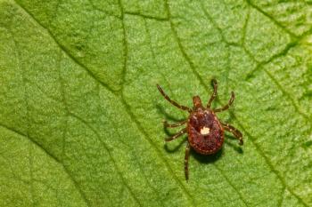
- Large silver spot at apex of scutum on females – hence name “lone star”
- All stages feed on wild & domestic animals, birds, & humans and is significant pest for humans & animals
- Can transmit Coxiella burnetii (Q-fever), tularemia, Ehrlichia chaffeensis, Ehrlichia ewingii, RMSF, Cytauxzoon felis (cats)
- Vectors agent of Southern Tick Associated Rash Infection (STARI) in humans
- Cause of STARI is currently unknown-may actually be the host reaction to tick saliva-leads to swelling and pain at bite region
Amblyomma maculatum (Gulf Coast tick)
- Southeastern US in Gulf coast region, but has expanded range recently
- Ornate scutum – often confused with Dermacentor-examine mouth parts to differentiae
- Adults attack nearly all animals & humans and can transmit Hepatozoon americanum Hepatozoonosis-dog must eat tick to be infected with Hepatozoon
All ixodes spp.
- Inornate ticks and No festoons, has distinct anal groove anterolateral to anal orifice
- Used for identification in NON-ENGORGED tick but can't see groove in engorged ticks-use mouth parts instead
Ixodes scapularis (black-legged tick)
- Wide distribution, in East, South, and Midwest U.S. Highest populations in upper Midwest and New England/midatlantic states
- Primary Lyme disease (Borrelia burgdorferi) vector in Eastern US and Midwest
- Vectors Babesia microti, Anaplasma phagocytophila
Ixodes pacificus (California black-legged tick)
- Primary Lyme disease vector in the West Coast
Tick-borne diseases
Tick paralysis
Potentially fatal reaction to a paralyzing neuromuscular toxin secreted in the saliva of a female tick late in her feeding. Cattle, sheep, horses, dogs, and humans seem to be most affected.
Clinical signs include: headache, vomiting, general malaise, loss of motor function and reflexes, followed by paralysis that starts in the lower body and spreads to the rest of the body.
Respiratory failure and death can result. Signs disappear rapidly when tick is removed, suggesting that the toxin is rapidly excreted or destroyed.
Lyme borreliosis
- Agent: Borrelia burgdorferi
- Vector: Ixodes scapularis (Eastern and Midwestern US), Ixodes pacificus (Western US)
- Geographical distribution: New England and mid-Atlantic states, upper Mid-west, and Pacific coast.
- Animal health: Major cause of canine and equine disease, including endocarditis and joint pain. Most cases occur in the spring and summer, during nymphal emergence, and in late fall and winter, during adult emergence.
- Human health: Acute and chronic diseases including joint pain, heart disease, and neurological disorders. Most cases occur in the spring and summer, during nymphal emergence, and in late fall and winter, during adult emergence.
- Diagnoses: Lyme disease is diagnosed using serology tests, bacterial cultures, and/or PCR of tissue (NOT BLOOD). Blood may be used for PCR in very acute cases, otherwise tissue biopsy is needed. Predictive value influences serological test interpretations-only treat animals with clinical signs suggestive of disease!!!
Rocky mountain spotted fever
- Agent: Rickettsia rickettsia
- Sometimes placed in “Spotted Fever” disease group
- Vector: Dermacentor variabilis
- Geographical distribution: Eastern US mainly. Most frequently reported tick borne disease in the eastern US. Animal health: Recent evidence has shown that untreated RMSF may lead to death of the affected animal. Clinical signs include whole body pain and are painful on palpation.
Cytauxzoon felis
Piroplasm of cats. Bobcats are reservoir host that is transmitted by Amblyomma americanum. Clinical signs: fever, dehydration, icterus, lymphadenomegaly, and hepatosplenomegaly. Treatment with atovaquone plus azithromycin. Diagnosis: PCR, blood smear (negative blood smear does not rule out infection) since early stage only see schizonts in macrophages. Prevention: Keep cats indoors!! Use preventative for tick infestation
Anaplasma phagocytophilum
- Intracellular rickettsia that causes human granulocytic anaplasmosis
- Infects granulocytes and leads to bleeding, fever, leukopenia,
- Clinical signs/symptoms may be worse with co-infection with Lyme or Babesia
- Vectored by Ixodes scapularis so same geographical distribution as Lyme Disease. Can be transmitted by blood transfusion.
- Diagnosis: clinical signs, PCR (acute cases), serology (chronic), CBC to look for leukopenia, Blood smear to look for morulae in granulocytes.
- Don't treat animals that are clinically normal but are only seropositive-potential false positive due to positive predictive value.
- Treatment with doxycycline or minocycline
Anaplasma platys
- Intracellular rickettsia that causes infectious cyclic thrombocytopenia in dogs
- Common clinical signs include bleeding, due to cyclic thrombocytopenia…may be worse with co-infection with Ehrlichia canis, which is transmitted by same tick.
- Transmitted by Rhipicephalus sanguineus –worldwide distribution
- Diagnosis: clinical signs, PCR (acute cases), serology (chronic). Don't treat animals that are clinically normal but are only seropositive-potential false positive due to positive predictive value.
- Treatment with doxycycline or minocycline
Ehrlichia canis
- Intracellular rickettsia that causes canine ehrlichosis
- Infects monocytes and leads to fever, anorexia, lethargy, thrombocytopenia, lymphadenopathy, edema, bone marrow suppression.
- The acute stage is mainly due to a vasculitis. E. canis replicates in monocytes. The infected monocytes bind to vascular endothelial cells and leads to a vasculitis
- Transmitted by Rhipicephalus sanguineus –worldwide distribution
- Diagnosis: clinical signs, PCR (acute cases), serology (chronic), CBC to look for leukopenia, Blood smear to look for morulae in monocytes
- Don't treat animals that are clinically normal but are only seropositive-potential false positive due to positive predictive value.
- Treatment with doxycycline or minocycline
Newsletter
From exam room tips to practice management insights, get trusted veterinary news delivered straight to your inbox—subscribe to dvm360.


