Troubleshooting dental radiography (Proceedings)
Dental radiographs are in essential part of the oral exam. The crown is just the tip of the iceberg. Approximately 42% of dental pathology is found subgingivally. Radiographs will help diagnose pathology that is not visible from the surface, confirm suspect pathology as well as help demonstrate the pathology to the client. Survey radiographs can also increase your clinic's revenue.
Dental radiographs are in essential part of the oral exam. The crown is just the tip of the iceberg. Approximately 42% of dental pathology is found subgingivally. Radiographs will help diagnose pathology that is not visible from the surface, confirm suspect pathology as well as help demonstrate the pathology to the client. Survey radiographs can also increase your clinic's revenue.

Table 1: Light Radiographs with Poor Contrast
Ideally, a full survey set of radiographs should be taken on all patients annually. This survey consists of six views, however on larger animals additional views may be required. Realistically, however, most clinics don't take full survey series. Radiographs should be taken when following problems are present: periodontal disease, endodontic disease, FORL's, draining tracts, trauma, oral masses, dental abnormalities and pre, intra and post surgical evaluations.
Dental radiograph units are relatively inexpensive. They are low maintenance and the film and chemicals are inexpensive. You can check with dental supply companies and purchase used units very reasonably. Medical radiograph machines can be used but are inconvenient and they do not show the detail necessary to make a definitive diagnosis. Dental radiograph units allow for accurate positioning without having to move the patient. They are compact, maneuverable, have limited settings and less radiation scatter. The settings for kVp and MA are pr]eset, leaving exposure time as the only adjustable setting.
Intraoral films (D speed/Ultraspeed™)can be used with the standard x-ray machines using a technique of 100mA, a focal length of 12-16 inches and an exposure time of 1/10th second. The kVp will vary from 50-85 dependent on patient size. As with standard radiographs, adjustments can be made if the dental radiograph is too light, increase the kVp or exposure time and if the radiograph is too dark, decrease the kVp or exposure time.

Table 2: Dark Radiographs with Poor Contrast
Chairside darkrooms are available that are inexpensive, provide rapid results and are easy to use. It is possible to purchase an automatic processor that is dental x-ray specific but they can be expensive. Holder clips are used to hold the film as you develop to avoid fingerprints. Light boxes with a magnifying lens are important to read the films.
Dental films come in four sizes with the most common sizes being 2 & 4. The film has a bubble on the upper left hand corner. This bubble should always be placed toward x-ray beam and towards to caudal aspect of the oral cavity to aid in orientation of the film. The film has multiple layers that include the white plastic outer layer, a silver lead layer, a paper layer and the film.
Dental radiographs need to be labeled, however due to the small size, the use of radio opaque markers or permanent markers may interfere with the radiographs. Cardboard or plastic holders are available that have space to label the radiographs. These holders can then be placed in the patient file or a specific storage cabinet. Envelopes and slide holders also maybe used successfully. I have found using a plastic business card holder that can be placed in a 3-ring binder are very useful for storing film size number 0, 1 & 2. A similar holder used for baseball cards works well for number 4 films.
As with any type of radiation, it is important to observe radiation safety guidelines. The amount of radiation needs to be kept to a minimum. If possible, step outside of the room but if it is not possible stay at least 6 feet away and out of the line of the beam. Always wear your film badge. There is a full range of positioning devices available to help keep the film in place. A gauze 4X4 works very well and is disposal and inexpensive.

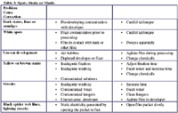
Table 3: Spots, Stains or Marks
A full radiographic survey will include 6 radiographs; anterior maxilla, anterior mandible, posterior maxilla (left & right), posterior mandible (left & right). There may need to be a need for additional views for specific teeth or in larger animals.
There are two intraoral radiograph techniques commonly utilized in veterinary dentistry. The simplest is the parallel technique and as luck would have it, it can be used for the fewest views. The parallel technique is used for the posterior mandible. This view will include the molars and caudal premolars. The film beam is placed at a 90 degree angle to the film, which has been placed on the lingual surface of the teeth.
The other technique is the bisecting angle. The bisecting angle is used to minimize distortions of the teeth. The bisecting angle is used for the anterior teeth, maxilla and mandible, the posterior maxilla teeth. In this technique the beam is aimed at the imaginary line bisecting the plane of the tooth and the plane of the film.
Bisecting Angle Parallel Technique
If the beam is not perpendicular to the bisecting angle the tooth will be distorted. The angle is too low it will cause elongation and too high it will cause foreshortening.
The maxillary P4 is a three-rooted tooth. If you use the bisecting angle technique the palatal root will be superimposed behind the mesiobuccal root. Using the SLOB rule will result in viewing all three roots. (Same Lingual, Opposite Buccal) The tube head is shifted slightly rostal or caudal to visualize all three roots. If the tube head is moved caudally, the palatal or lingual root will be most caudal on the radiograph. If the tube head is moved rostrally, the lingual root will be the most rostral root on the radiograph.
Standard bisecting angle:
D P M
SLOB Rule tubehead caudal
D P
M
SLOB Rule tubehead rostral
M P
D
In cats, radiographs of the maxillary premolars and molars utilizing the bisecting angle technique results in superimposition of the zygomatic arch or the apex of the root. To eliminate this superimposition, an extraoral view of the feline maxillary premolars and molar may be used. You may also intentionally elongate the view to eliminate the superimposition.
To view the radiographs, orient the film on the viewbox so that the raised dot is projecting toward you. The resultant image is as if you were looking from the outside of the mouth inward.
The goal of dental radiology is to take diagnostic radiographs. It is important to have the correct teeth in your radiographs as well as the entire tooth with a minimum of 2 mm of bone around the roots. Contrast is vital, avoid over or under exposing the film. Films that are underexposed will have a white appearance, the teeth and bones seem to coalesce so it makes it difficult to determine one from the other. Overexposure of the film will give a dark film, lack contrast and have a ghost like appearance. Cervical burnout or blackening of the neck of the tooth can happen in overexposure.
Technical errors can occur at any stage in dental radiology. This can be due to film placement, patient positioning, angle of the x-ray beam, exposure, processing, storage or any combinations of the above. The tables below addresses the most common problems you will encounter.
Interpretation of the radiographic signs is beyond the scope of this lab. There are several excellent veterinary dental radiograph textbooks available to assist in interpretation.
References
1.Mulligan, TW, Aller, MS, Williams, CA Atlas of Canine & Feline Dental Radiography. Vet. Learning Systems, 1998.
2.Holmstrom, SE, Frost, P, Gammon, RL Veterinary Dental Techniques for the Small Animal Practitioner. WB Sauders, Co. 1992
3. Holmstrom, SE, Veterinary Dentistry for the Technician and Office Staff, W.B. Saunders, 2000.