UC-Davis shows vitamin E role in treating equine neurologic disease
Horses' condition makes it easier for vitamin E to do its work protecting cells from free-radical damage.
Some inflammatory or degenerative neurologic diseases that affect horses — equine degenerative myeloencephalopathy (EDM) and equine motor neuron disease (EMND) — are linked to vitamin E deficiency. Vitamin E is a dietary antioxidant that protects cell membranes and tissues from oxidant free-radical damage, which is believed to play a role in the etiology and progression of the diseases.

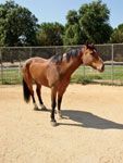
Photo 1: A horse with EMND showing postural muscle weakness with generalized muscle wasting. Note the position of the horse's hind legs.
Now, researchers at the University of California-Davis say horses with neurologic disease may respond differently to vitamin E treatment because of disruption of the blood-brain barrier or increased oxidant damage associated with the underlying disease.
The blood-brain barrier of horses with neurologic disease may be potentially compromised, allowing vitamin E to cross more easily, show higher cerebrospinal fluid (CSF) concentrations and be more effective in reducing oxidant free-radical damage in the brain.

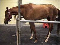
Photo 2: A horse with EDM shows proprioceptive deficits.
Conversely, vitamin E may be more readily metabolized due to increased oxidant damage, and therefore show lower CSF values at the desired site of action. The antioxidant "demand" for older horses might be greater, especially those with neurologic disease.
The researchers demonstrated that daily oral administration of 10,000 IU vitamin E, as micellized d-alpha-tocopherol, crosses the blood-brain barrier in healthy horses. Leading the work were Jamie Higgins, DVM, Dipl. ACVIM, fellow in large-animal emergency medicine and critical care in the UC-Davis School of Veterinary Medicine, and colleagues, including Nicola Pusterla, DVM, Dipl. ACVIM, associate professor in equine internal medicine.
Vitamin E may benefit horses with neurologic disease, the researchers say. CSF samples they collected from the atlanto-occipital space provided accurate values of alpha-tocopherol concentrations around the brain and cervical portion of the spinal cord, which is where the most damage to the central nervous system is detected in horses.
They found that serum alpha-tocopherol concentration increases rapidly in a nonlinear manner if 10,000 IU of d-alpha-tocopherol supplement is given orally daily, compared to 1,000 IU.
Serum alpha-tocopherol concentrations in both groups increased significantly throughout the period of vitamin E administration. From baseline values at Day 0, they reached a plateau by Day 10 (mean, 2.96 µg/ml, 1,000 IU; 6.6 µg/ml, 10,000 IU).
Median and maximum CSF alpha-tocopherol concentrations were higher in horses treated with 10,000 IU d-alpha-tocopherol, compared with those receiving 1,000 IU, suggesting that high doses of supplemental vitamin E are likely of greater antioxidant benefit.
Both groups within 10 days showed a 1.3- to 3.4-fold increase in CSF alpha-tocopherol in nine out of 10 horses.
In another study, Pusterla, Higgins and colleagues showed that feeding of natural, micellized d-alpha-tocopherol showed greater bioavailability than synthetic (d,l-alpha-tocopherol), when fed on an equal basis to produce greater plasma and CSF alpha-tocopherol levels.
It was concluded that, because maintenance of sufficient cerebral alpha-tocopherol concentrations is essential for neurologic function, and of even greater importance when brain function is impaired by degenerative or inflammatory neurologic disorders, supplementation of a micellized alpha-tocopherol should be used preferentially over synthetic vitamin E when treating horses with neurologic disorders.
Horses presented to UC-Davis with neurologic disease routinely get 10,000 IU daily of alpha-tocopherol.
EDM diagnosis, treatment
Equine degenerative myeloencephalopathy (EDM) is a diffuse degenerative disease of the spinal cord and brain stem, with signs of slowly progressive ataxia and weakness. Though first observed in the Northeast, several breeds of horses with ataxia and neuroaxonal dystrophy were subsequently noted throughout the United States, including Appaloosas, Arabians, Quarterhorses, Thoroughbreds, Standardbreds, Paso Fino horses, Morgan horses, Paint horses, Norwegian Fjord horses, Welsh ponies and mules.
The disease also was seen in Grant zebras (E. burchelli) and Mongolian wild horses (E. przewalskii) in captivity. Horses with ataxia and the neuronal dystrophy characteristic of EDM as well as atactic wild horses and zebras responsive to therapy with vitamin E have been identified in Europe. Most EDM-affected horses exhibit ataxia during their first year, with onset of clinical signs prior to 6 months of age. The initial clinical signs were upper motor neuron signs with general proprioceptive deficits as well as a tendency for generalized hyporeflexia and hypometria. Signs of EDM include clumsiness, inability to do complicated movements, malpositioning of limbs at rest and during movement and obvious ataxia or incoordination.
EDM, commonly seen in young horses, was shown to have a familial component, but is also linked to low serum vitamin E levels and/or vitamin E deficiency.
Several factors suggest vitamin E deficiency. Pathologic findings of neuroaxonal dystrophy and lipofuscin-like pigment accumulation are common features in experimentally induced vitamin E deficiency in several species and in EDM-affected horses.
Low to deficient serum or plasma alpha-tocopherol values were reported in several studies of EDM. Supplementary administration of vitamin E to foals from sires known to produce EDM decreased the incidence of the disease, and adult horses with EDM treated with 6,000 IU/day of alpha-tocopheryl acetate for one or more years showed neurologic improvement.
It was found that foals showing signs of incoordination, supplemented with 6,000 IU vitamin E per day appeared normal by 2 years of age. (The EDM work is by Linda Blythe, DVM, PhD, and Morrie Craig, PhD, researchers at Oregon State University, 1991-1992.)
It is highly recommended that horses at risk be supplemented with the vitamin. The incidence of EDM on one farm was reduced from 40 percent to zero via supplementation of 1,000 IU to 2,000 IU per day of d,l alpha-tocopheryl acetate. Horses fed 6,000 IU of d,l alpha-tocopheryl acetate per day, mixed with 60 ml of corn oil, mixed in a liter of sweet feed or grain, showed improved neurologic scores.
In children with neurologic deficits caused by malabsorption of vitamin E, therapy with massive doses of vitamin E before 5 years of age resulted in return to normalcy. Young horses respond better to vitamin E supplementation if their gait deficits are appreciated sooner, the UC researchers say.
The reasons for the low alpha-tocopherol values may include inadequate vitamin E in the feed; reduced absorption of vitamin E due to lack of bile acids or intestinal malfunction; failure to make chylomicrons in the enterocytes necessary to carry the vitamin E via the lymphatic system to the blood and liver; the absence of specific hepatic protein (tocopherol transfer protein, TPP) postulated to transfer the vitamin E from the chylomicrons to the LDL and HDL; an inadequate number of cellular vitamin E receptors; and increased utilization and/or excretion of the vitamin.
Vitamin E is valued for its prime role as a scavenger of free-radicals and therefore its antioxidant protection of neuronal lipid membranes. The disease often is confirmed, not only by the ataxia seen in young horses, but also by low plasma or serum α-tocopherol status (< 3 µg/ml), though these low values also are seen in horses without neurologic disease. A known history of EDM within the horse's bloodline can help confirm the diagnosis.
Supplementing stallions with 1,500 IU vitamin E per day decreases the incidence of EDM in their foals from 40 percent to 10 percent. A vitamin-E-responsive EDM in Standardbred and Paso Fino horses from 3 to 30 months of age has been described. Symmetric ataxia and paresis, along with laryngeal adductor, cervicofacial, local cervical and cutaneous trunci hyporeflexia, were characteristic. No clinical signs were observed before 3 months of age.
The onset of gait abnormality is usually abrupt. Clinical signs have been seen to remain static or progressed for weeks or months. Severely affected animals often fall while running. In one study, the mean (± SD) serum alpha-tocopherol concentration of 13 ataxic weanlings was 0.62 ± 0.13 (range 0.47 to 0.84) µg/ml.
A number of non-ataxic weanlings had similar values, although vitamin E supplementation reduced the incidence of the syndrome on affected farms. Based on genetic studies, this disorder seems to have a familial disposition.
To reach a tentative diagnosis of EDM, a neurologic exam and a CSF exam are recommended. Measuring various parameters within the CSF are suggested, and analysis of CSF alpha-tocopherol may be of value as shown by the results of a 2008 study by Higgins and colleagues.
EMND diagnosis, treatment
Equine motor neuron disease (EMND) is a naturally occurring neurodegenerative disease, an oxidative disorder of the somatic motor neuron system in adult horses.
Clinical signs include weight loss from muscle wasting, trembling, muscle fasciculations and prolonged periods of recumbency. Pathologic changes are primarily limited to the somatic lower motor neurons of the spinal cord and brainstem nuclei, and their corresponding efferent nerves and innervated muscle groups and the retinal pigment epithelium.
Vitamin E deficiency is a primary factor, as studies show that horses without access to green forage/pasture for 18 months or more, high-grain diets and coprophagia and/or geophagia are at high risk for the disease.
Low plasma/serum alpha-tocopherol is a consistent laboratory finding in horses with EMND, along with increased amounts of potential pro-oxidants, copper in the spinal cord and of hepatic iron.
The lack of access to pasture, dietary deficiency of vitamin E, or excessive dietary copper have been confirmed as likely risk factors for the disease.
Four of eight horses fed low-vitamin-E diets (hay stored for > 1 yr. 19.8 IU/kg; grain, 25.7 IU/kg) with excess copper (> 4,000 ppm) and iron (> 2,000 ppm) exhibited signs of EMND at 21, 27 and 28 months, with mean plasma alpha-tocopherol concentrations of 0.255 µg/ml. (EMND work is by Thomas J. Divers, DVM, Dipl. ACVIM, Dipl. AVECC, professor of medicine and chief of the Section of Large Animal Medicine at Cornell University College of Veterinary Medicine, and colleagues, 2006).
The other four horses, though they did not show signs of EMND within 30 months, did show decreased plasma alpha-tocopherol levels (mean, 0.39 µg/ml). Histological changes in the spinal cord, peripheral nerves and muscles were characteristic of EMND in all four affected horses. The other four horses, though they had no clinical signs of EMND, did show some nerve lesions histologically. Mean hepatic alpha-tocopherol concentration was significantly low (2.57 ug/g, dry weight) in EMND affected horses compared to control horses (21.1 ug/g).
Mean hepatic copper concentrations (503 ppm) of horses with EMND were excessively higher, more than 50 times the value of control horses.
It was concluded that vitamin E deficiency plays a major causative role in EMND. Vitamin E was not measured in the spinal cord tissue of the horses, but in humans tocopherol concentrations in CSF are highly correlated with serum concentrations.
Podcast CE: A Surgeon’s Perspective on Current Trends for the Management of Osteoarthritis, Part 1
May 17th 2024David L. Dycus, DVM, MS, CCRP, DACVS joins Adam Christman, DVM, MBA, to discuss a proactive approach to the diagnosis of osteoarthritis and the best tools for general practice.
Listen