Ultrasound of arterial and venous thrombosis
Thrombosis is a complication of many diseases in veterinary medicine.
Thrombosis is a complication of many diseases in veterinary medicine.
Heart disease, protein-losing nephropathy and steroid therapy or hyperadrenocorticism can all predispose an animal to arterial or venous thrombi. Many of the systemic vessels involved are located in the abdomen and visible on abdominal ultrasound. We can identify thrombosis during an acute episode or as an incidental finding. Here are some of the features you might see:
Appearance of the thrombus
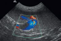
In the acute phase, thrombi in the arterial or venous systems typically are anechoic. These can be caused by migration of a fragment of thrombus from the left atrium to the terminal aorta (such as cats with cardiomyopathy), or a portal vein thrombus that causes portal hypertension and ascites. You may see some faint echogenicity within the vessel, but these usually are diagnosed using Doppler ultrasound. The color flows around the filling defect in the vessel.

Image 1
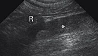
After several days, the thrombus organizes into a visible structure. It is usually intermediate in echogenicity, and can partially or completely fill the vessel (Image 1). An older thrombus may contract so there is flow around it (Image 2).

Image 2
Location of the thrombus
The location of the thrombus determines whether it causes clinical signs. Arterial thrombi, such as in the aorta, can occlude blood flow to distal structures. Ischemia of the hind limbs is a common complication of aortic thrombosis, because the thrombus can occlude or extend down the iliac arteries.
An aortic thrombus also can occlude the renal arteries, causing renal ischemia. The thrombus in Image 3 (*) is very close to the renal artery (R) but not occluding it. Portal-vein thrombosis also is usually clinical because of the ascites that forms from portal hypertension.

Image 3
Thrombi in the splenic veins are very common and may not cause any symptoms. These can be an incidental finding (Images 1 and 2), such as in this dog with lymphoma that had been treated with prednisone.
Tumor thrombus
Tumors can cause physical obstruction of a vessel, usually in the caudal vena cava. Any tumor with a tributary joining the caudal vena cava can infiltrate the vessel and reach the systemic venous circulation.
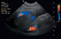
The most common tumor that invades the caudal vena cava is an adrenal tumor (Image 4), with pheochromocytomas having a predisposition. The tumor thrombus starts by traveling down the phrenicoabdominal vein that runs across the mid-portion of the gland, then reaches the caudal vena cava. Renal tumors can follow a similar pattern and invade down the renal vein.

Image 4
How to evaluate thrombosis:
» Use color Doppler to look for acute thrombi.
» Evaluate the extent and location of visible thrombi.
» Check for peripheral flow with color Doppler.
» Look for evidence of neoplasia in the region.
» Check for sequelae of thrombosis such as ischemia of distal structures or ascites.
Dr. Zwingenberger is a veterinary radiologist at the University of California-Davis.
Veterinary scene down under: Australia welcomes first mobile CT scanner, and more news
June 25th 2024Updates on the launch of the first mobile CT scanner available for Australian pets; and learn about the innovative device which simplifies placement of urinary catheters in female dogs
Read More