Update on babesiosis, leptospirosis in dogs
Q: Please provide a review on canine babesiosis and canine leptospirosis.
Q: Please provide a review on canine babesiosis and canine leptospirosis.
A. Dr. Douglass K. Macintire lectured on Diagnosis and Treatment of Canine Babesiosis and Canine Leptospirosis at the 2007 American College of Veterinary Internal Medicine Forum in Seattle. Here are some relevant points from his presentation:
Canine babesiosis is an emerging infectious disease in North America. Historically, the disease has been classified either as "large babesia" (B. canis) or "small babesia" (B. gibsoni) on the basis of the size of intraerythrocytic forms. There are three subspecies of the "large" babesia: B. canis vogeli (found in the United States), B. canis canis (found in Europe) and B. canis rossi (found in South Africa). Large babesia piroplasms appear as bi-lobed, pear-shaped organisms in the red blood cells.
There are at least three genetically distinct, but morphologically indistinguishable, small piroplasms that can infect dogs. Babesia gibsoni was first diagnosed in Asian dogs but now is reported worldwide. It is being diagnosed in epidemic proportions in American Pit Bull Terriers throughout the United States. A second small babesia was reported in dogs from California in 1991. Although the organism was initially thought to be B. gibsoni, subsequent PCR analysis has reclassified the piroplasm as a Theilena species. A third small piroplasm, a babesia microti-like organism has been found in dogs in northwest Spain as a newly emerging pathogen.
Johnny D. Hoskins DVM, PhD, Dipl. ACVIM
Babesiosis can cause severe, life-threatening signs in some dogs, while others show no outward signs of disease. Common findings in affected dogs include hemolytic anemia, thrombocytopenia, hemoglobinuria, icterus, pallor, splenomegaly, lymphadenopathy and fever. Clinical signs resemble those associated with immune-mediated hemolytic anemia, immune-mediated thrombocytopenia or other tick-borne diseases. It is important to identify the correct diagnosis, as early recognition and appropriate treatment are important for reducing associated morbidity and mortality.
Transmission, diagnosis
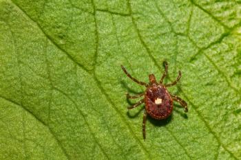
The vectors known to transmit babesiosis include Rhipicephalus species, Haemaphysalis species and Dermacentor species. The tick must feed for two to three days for transmission to occur from the salivary glands of the infected tick to the dog's bloodstream. The parasites enter red blood cells, causing extravascular and intravascular hemolysis. Autoagglutination and secondary immune-mediated destruction of red blood cells may occur.
Diagnosis can be confirmed by finding the organism on a blood smear or by PCR testing. Only the PCR test can identify species or subspecies, and it is more sensitive in detecting subclinical carrier animals than a blood smear. Serologic titers >1:64 are considered positive, but they do not differentiate clinical exposure from active infection, and there is cross-reactivity between species. Dogs with a positive babesia titer should not be used as blood donors.
Correct diagnosis is key: Babesiosis can cause severe, life-threatening signs in some dogs, while others show no outward signs. That's why early, correct identification of the disease is important.
Other means of transmission besides arthropod vectors include blood contamination through poor kennel practices, such as sharing needles or surgical instruments, dog fighting and blood transfusions. Vertical transmission from dam to offspring is suspected as a means of transmission among American Pit Bull Terriers and Greyhounds. About 50 percent of American Pit Bull Terriers surveyed have a positive PCR test for B. gibsoni, and most have subclinical infections.
Treatment is recommended even in subclinically infected dogs to reduce the natural reservoir for transmitting the disease to arthropod populations. Aggressive control measures to eradicate the vector are key to eliminating spread of the disease.
The most commonly used treatment is imidocarb dipropionate, 6.6 mg/kg IM or SC once, repeated in seven to 14 days. Pretreatment with atropine (0.04 mg/kg IM or SC 30 minutes prior to imidocarb) is recommended to decrease cholinergic side effects. Supportive care with a blood transfusion or Oxyglobin® (10-30 ml/kg IV) may be needed in severely anemic dogs.
Dogs that are febrile and dehydrated may require intravenous fluid support. Treatment with imidocarb usually does not eliminate the parasite, but most dogs show marked clinical improvement. Recently, a new treatment has shown promise in eliminating the organism, but it is very expensive. Dogs treated with atovaquone (13.4 mg/kg PO q 8 h with a fatty meal) and azithromycin (10 mg/kg PO q 24 h) for 10 days became negative on PCR testing 60 to 90 days post-treatment. If anemia and thrombocytopenia do not improve with anti-protozoal treatment, immunosuppressive therapy with prednisone (1 mg/kg q 12 h) may be needed and tapered off gradually until anemia resolves.
The babesia microti-like organism in Spain has been associated with azotemia and protein-losing nephropathy. The cause of renal pathology is thought to be secondary to immunoproliferative glomerulonephritis, but more research is needed to better elucidate the renal lesions.
Canine leptospirosis
Leptospirosis is caused by the spirochete bacteria Leptospira interrogans sensu lato. More than 150 distinct serovars have been identified, and eight are known to infect dogs. Leptospira serovars infect 160 mammalian species. Maintenance hosts are carrier animals that provide a reservoir for the bacteria. Incidental hosts are other animals that have a more severe clinical illness and a briefer period of shedding. Maintenance hosts for the different serovars include dogs (canicola); rats (icterohemorrhagiae); raccoons, skunks and opossums (grippotyphosa); cattle and pigs (pomona); pigs (bratislava); cattle (hardjo); and mice (ballum).
The most common serovars infecting dogs in the United States used to be icterohemorrhagiae and canicola. After widespread use of vaccines containing these serovars, the incidence of classic leptospirosis caused by these serovars decreased. There is no cross-protection against other serovars, and in recent years grippotyphosa, pomona, autumnalis and bratislava have emerged as more common pathogens causing leptospirosis in dogs.
Animals become infected with leptospirosis through direct contact with infected urine, ingestion of infected tissues or exposure from animal bites. Indirect transmission occurs through contact with contaminated water, soil, bedding or food. Most cases of leptospirosis occur in late summer and early fall, especially after heavy rains or flooding. Exposure to stagnant water contaminated by wildlife urine appears to be a major source of infection for dogs.
The spirochete bacteria penetrate abraded skin or mucous membranes and multiply in the blood and other tissues, causing hepatic and/or renal disease and vasculitis. Herding dogs, hounds, working dogs and male dogs are at increased risk. Serovar icterohemorrhagiae causes icterus, liver failure, uremia and pyrexia. The canicola serovar causes acute nephritis with less hepatic involvement, and serovars pomona, bratislava, autumnalis and grippotyphosa cause acute renal failure without hepatic insufficiency.
Clinical signs
Clinical signs of leptospirosis include lethargy, depression, anorexia, vomiting, dehydration and fever. Renomegaly and abdominal pain also are common. Some dogs have CNS involvement, including neck pain, ataxia or seizures. Icterus may be present in dogs with hepatic involvement.
Leukocytosis and thrombocytopenia are the most common abnormalities on the hemogram. Abnormalities in serum chemistries include azotemia, hyperphosphatemia, metabolic acidosis and elevated liver enzymes. Vomiting often causes hyponatremia, hypochloremia and hypokalemia, but hyperkalemia may be present in severe cases with acute renal failure. Urinalysis reveals casts, renal tubular epithelial cells, blood cells, glucosuria, proteinuria and bilirubinuria. Specific gravity is usually <1.020. Darkfield microscopy of the urine may reveal spirochetes, but these cannot be differentiated from Borrelia spirochetes. Because of periodic shedding, a negative darkfield microscopy does not rule out leptospirosis.
Thoracic radiographs may reveal a reticulonodular pattern with a caudo-dorsal distribution. These pulmonary changes are thought to be secondary to vasculitis and/or pulmonary hemorrhage. Abdominal ultrasound study often reveals bilateral renomegaly with hyperechogenicity of the renal cortices. There may be a medullary band of hyperechogenicity and dilation of the renal pelvises.
The microscopic agglutination test (MAT) is the most common serologic test employed for diagnosis. Multiple serovars are tested, and a single titer >1:800 associated with clinical signs is considered diagnostic. There is some cross reaction between serovars, and when several are positive, the one with the highest titer is considered to be the infective agent. Vaccination titers usually are low, ranging from 1:100 to 1:400. In acute disease, titers may be negative for seven to 10 days, and retesting in two to four weeks is recommended. Antibiotics will lower the magnitude of the titer.
An ELISA test is available for IgM and IgG antibodies, but does not delineate serovars or vaccine titers. The IgM titer is increased seven to 14 days post-infection, and the IgG titer is elevated two to three weeks post-infection.
A PCR test is available through some laboratories. It is especially useful for identifying early infections and carriers, and is considered sensitive and specific. Other methods of diagnosis include silver stains or immunohistochemistry of renal or hepatic biopsies with identification of the spirochetes on histopathologic sampling.
Treatment and precautions
Treatment of leptospirosis involves antibiotic therapy and supportive care for acute renal failure. Early antibiotic therapy inhibits multiplication of the organism and limits the hepatic and renal damage. Initial antibiotics of choice are either penicillin G (25,000-40,000 U/kg SC, IM, or IV q 8 h) or ampicillin (22 mg/kg PO, SC, or IV q 8 h) for 14 days to eliminate leptospiremia. A second course of antibiotics is required to eliminate the carrier state. This is accomplished by administering doxycycline at a dosage of 5 mg/kg PO q 12 h for 2 weeks.
Because leptospirosis is a zoonotic disease, the following precautions should be taken when handling suspect cases:
- 1. Gloves, goggles and gown should be worn when handling the dog or hosing down runs.
- 2. The dog should be placed in a bottom cage with disposable bedding and not moved until it has received antibiotic therapy for three days.
- 3. Catheters, bedding, gloves and anything else that contacts the dog should be disposed of in a biohazard container.
- 4. If the animal urinates outside the cage, the area must be disinfected. Iodophors are effective in killing spirochete bacteria.
- 5. Hands should be washed immediately after handling the dog.
- 6. After discharge, the cage should be thoroughly cleaned and disinfected and not used for 24 hours.
Supportive care
Supportive care for acute renal failure involves the following recommendations:
- 1. Placement of a jugular catheter allows for frequent blood sampling, IV fluid diuresis and measurement of central venous pressure.
- 2. Placement of a urinary catheter allows for safe collection and disposal of urine through a closed system and monitoring of urine output in animals with oliguria (<1 ml/kg/h).
- 3. Fluid deficits are corrected rapidly over four to six hours using the following formula: percent dehydration x body weight (kg) x 1000 = number of ml. In addition, maintenance needs (1 ml/lb/h) and fluid to replace contemporary losses from vomiting and diarrhea are added to the hourly rate.
- 4. If possible, serum electrolytes and blood gases should be checked. Hyperkalemia can be treated with the following therapeutic options: (a) regular insulin (0.5 units/kg IV) plus 1 gram of dextrose per unit of insulin diluted to 20 percent and given IV; (b) sodium bicarbonate 1-2 mEq/kg slowly IV; or (c) calcium gluconate, 0.5-1 ml/kg IV over 10 to 15 minutes. Severe metabolic acidosis can be corrected with the following formula: 0.3 x body weight (kg) x base deficit = number of mEq NaHCO3 needed to correct the deficit. One-fourth to one-third of the dose is given slowly introvenously and the rest is added to the IV fluids.
- 5. After the fluid challenge, the animal's blood pressure should be normal to slightly high, and the CVP should be 5-8 cm H2O. If the urine output is < 1-2 ml/kg/h, polyuria should be induced by administering one or all of the following: (1) manni-tol, 0.5 g/kg IV over 20 minutes; (2) furosemide 2 mg/kg IV with repeated doses if no response seen or CRI of 3-8 ug/kg/min; (3) dopamine 0.5-3 ug/kg/min CRI.
- 6. After volume expansion has been achieved, the IV fluid rate is established by determining "ins and outs." Urine output is quantified every four hours. The new hourly rate is set by dividing the urine amount by four and adding 1 ml/lb for maintenance needs.
- 7. Nausea and vomiting are treated with metoclopramide (1-2 mg/kg/day CRI or 0.2-0.4 mg/kg q 8 h SQ). Gastric hyperacidity is treated with ranitidine (1-2 mg/kg q 12 h IV) or famotidine (0.5-1 mg/kg q 24 h IV) or sucralfate (0.5-1 gm PO q 8 h).
- 8. Nutrition can be provided by PPN, TPN or enteral feedings. Pancreatitis can be a serious complication of acute renal failure and should be ruled out prior to feeding. A restricted protein diet low in phosphorus is recommended.
- 9. Animals that do not respond to aggressive medical management may require hemodialysis or peritoneal dialysis.
- 10. Prognosis for recovery is fair. About 80 percent of dogs treated aggressively for leptospirosis recover within seven to 10 days. Some experience full recovery and some have residual renal insufficiency. Renal function should be monitored for the next six to 12 months.
A new vaccine is available that protects against serovars pomona and grippotyphosa as well as icterohemorrhagiae and canicola.
Dr. Hoskins is owner of Docu-Tech Services. He is a diplomate of the American College of Veterinary Internal Medicine with specialities in small animal pediatrics. He can be reached at (225) 955-3252, fax: (214) 242-2200 or e-mail:
Newsletter
From exam room tips to practice management insights, get trusted veterinary news delivered straight to your inbox—subscribe to dvm360.


