Urinary incontinence in dogs -- diagnosis and treatment (Proceedings)
Primary sphincter mechanism incompetence (idiopathic incontinence, hormone-responsive incontinence) is the most common and important acquired cause of incontinence in dogs.
Primary sphincter mechanism incompetence (PSMI)
PSMI (idiopathic incontinence, hormone-responsive incontinence) is the most common and important acquired cause of incontinence in dogs. It largely is a condition of spayed female dogs, but in some breeds incontinence may precede ovariohysterectomy (OHE). Decreases in maximal urethral closure pressure and functional urethral length predictably occur during the first 12 to 18 months after spaying, resulting in a caudal shift of the urethral profile, and a deterioration of urethral closure function. It is speculated that this decline in urethral closure pressure continues with advancing age.
Approximately 20% of female dogs will develop PSMI a mean of 2.9 years after OHE performed after their first heat (immediately to 12 years later). Dogs greater than 20 kg (31%) are more likely to develop PSMI than are those less than 20 kg (9.3%). Incontinence is about one-half as frequent in bitches that undergo OHE before their first heat, but episodes of incontinence are worse. In one study, early-age gonadectomy was associated with an increased rate of urinary incontinence prompting the recommendation that gonadectomy should be delayed until at least 3 months of age. In another study, female dogs neutered at 7 weeks of age did not have increased urinary incontinence compared to those neutered at 7 months of age. Boxers in Europe comprise 65% of cases whereas Dobermans and Giant Schnauzers predominate in the USA. Confirmation of the diagnosis of PSMI is made by finding low maximal urethral closure pressure and decreased functional profile length during urethral pressure studies.
Phenylpropanolamine (PPA) at 1.0 to 1.5 mg/kg PO BID to TID effectively controls incontinence in about 74 to 92% of dogs with PSMI by stimulating alpha-adrenoreceptors in the urethra and increasing urethral tone. Of those not completely continent following PPA, many will have some improvement in their {Kyles, 1998 #6}continence. OVER HALF of dogs treated with regular PPA that failed to respond, became continent when treated with sustained release PPA. The effect of PPA to control continence becomes less over time in some dogs. PPA sometimes must be given two to three times per day to control incontinence. Not all alpha agonists have the same effect as PPA had a greater effect than pseudoephedrine in a recent study. If incontinence only occurs during the sleeping hours, the highest dose can be given before bedtime. Rarely, some dogs display restlessness and mild behavioral changes on PPA which makes its use less attractive. The beneficial effect of alpha-adrenoreceptor stimulation appears to be greatest in dogs that were older and had OHE performed long before incontinence started. Dogs with systemic hypertension should not be treated with alpha-adrenergic agents that could aggravate their systemic hypertension. Similarly, dogs with clinically relevant cardiac or renal disease should not be treated with alpha-adrenergic agonists. Systemic blood pressure should be measured at baseline, one week, 1 month and at 3 months to ensure that hypertension is not emerging. Twice yearly measurements of systemic blood pressure are recommended thereafter.
Estrogens may be effective in controlling the incontinence of PSMI by increasing the number or sensitivity of alpha-adrenoreceptors in the urethra. Estrogens may have other less well-understood effects including increased urethral tone arising from vascular changes and reduction in circulating FSH and LH. Estriol increases urethral resistance in sexually intact and spayed female dogs without urinary incontinence. Estrogens relieve incontinence in PSMI in 65-83% of treated dogs. Diethylstilbesterol (DES) is available from compounding pharmacies, but it was removed from the general market due to medical concerns in human patients. DES at 0.5 to 1.0 mg per dog (0.02 mg/kg; maximal dose of 1 mg) for 3 to 5 days as an induction dose often is effective in reducing incontinence attributed to PSMI. The dose then is periodically decreased to every other day and then to the lowest dose that will maintain continence. Some dogs cannot tolerate DES as the dose required to maintain continence may induce clinical signs of estrus. Conjugated estrogens such as Premarin® are more readily available than DES and can be given at 20 micrograms per kg every 4 days as an alternative. Some pharmacists prefer the brand name drug (Premarin®) over generics because of bioequivalence issues. There is no published data on safety, but experience in the veterinary community suggests that this treatment is safe and effective in many cases. Bone marrow toxicity is a potential adverse effect of estrogen treatment, but treatment with low doses of DES or conjugated estrogens appears to be quite safe. Bone marrow hypoplasia has been observed with higher dose regimens of DES and most commonly when estradiol cypionate (ECP) was given. ECP should never be used as a treatment for incontinence - it is too dangerous. Intermittent low dose maintenance of DES or conjugated estrogen to control incontinence may be preferred over the multiple daily doses of PPA that must be given, despite the fact that PPA is more often effective in the control of incontinence. In difficult cases, DES can be used simultaneously with PPA to achieve a synergistic effect which may effectively control incontinence. In dogs with incontinence refractory to these medial treatments, detrusor instability (hyperactive bladder) may be contributing to the incontinence. A therapeutic trial with propantheline, oxybutinin, or flavoxate to relax the detrusor muscle may be warranted in these instances.
Treatment with gonadotropin-releasing hormone (GnRH) analogues recently has been reported to result in complete continence in over half of dogs with PSMI that failed traditional medical therapy; most of the remaining dogs were improved but not completely continent. An average of 253 days of continence was observed in the 7 dogs that became fully continent with GnRH as the sole treatment. All 5 dogs that were partially improved by GnRH treatment became fully continent when PPA treatment was added later. Treatment with GnRH analogues reduces the concentrations of FSH and LH that develop after OHE in female dogs. Increased FSH and LH either directly or indirectly may play a role in development of PSMI in susceptible dogs. However, maximal urethral closure pressure does not appear to be directly related to FSH or LH concentrations. Initial treatment with GnRH analogues instead of conventional medical therapy was provided for two dogs of this series. Long acting GnRH analogues are effective as a first-line treatment for PSMI with a success rate of 71%, but this is lower than that achieved with alpha-adrenergics. Treatment with GnRH (leuprolide) did not increase urethral closure pressure despite gaining urinary continence. Receptors for GnRH, FSH, and LH have been demonstrated in various regions and densities in the urethra and bladder of the dog.
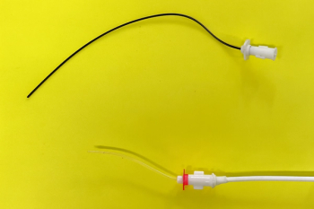
Urethral bulking agents usually are used in dogs that have failed medical therapy for PSMI. Periurethral submucosal injections of these agents are administered through a cystoscope. Other candidates for urethral bulking are dogs that do cannot tolerate alpha-adrenergic drugs or estrogens. Initial reports showed a control rate of 53% for incontinence treated with one or two series of injections of medical grade collagen. The rate of continence was 75% when PPA was added to dogs in which the injections provided inadequate urinary control. Recent improvements in technique suggest an 80% success rate with one or two injections and a 90% success rate if PPA is added to dogs that initially failed; 10% are not helped despite injections and drug therapy. A success rate of 68% in 40 dogs treated with submucosal collagen injections recently was reported to last a mean of 17 months. In a review of collagen injections at our institution, complete continence was achieved by many dogs, and nearly all dogs improved markedly in their degree of continence for many months based on scoring records completed by owners. Some dogs require a second series of injections, the second series of injections usually is easier to complete because the previous urethral bulking site is readily identified and augmented. Three submucosal injections of 0.5 to 1.0 ml of collagen are administered in the proximal urethra while the bladder is moderately distended with fluid so as to put some tension on the urethra. The amount of collagen injected is determined visually. The goal is for the 3 submucosal urethral protrusions to touch one another in the center of the urethral lumen. About one-third of the injected collagen is absorbed from the phosphate-buffered saline in the commercial preparation.
Figure A,B,C. Injector needle is advanced to urethral site through a cystoscope. Injection of collagen is given about 0.5 to 1.0 ml per injection site. Ideally, the submucosal injections provide a bulking effect with appostion from the various injection sites.
Ectopic ureters
Ectopic ureters are the most common congenital cause for urinary incontinence, and occur primarily in female dogs of high risk breeds (e.g., Golden Retriever, Labrador Retrievers, Siberian Husky, soft-coated Wheaten Terrier). Male dogs with ectopic ureters rarely exhibit signs of incontinence, perhaps as a result of a much longer distal urethra that could allow for continence despite more proximal termination of the ectopic ureter. Urinary incontinence usually is nearly constant and may be associated with intermittent or persistent bacterial urinary tract infection. The history of urinary incontinence usually can be traced to the time of weaning and often initially is mistaken as a behavioral problem associated with housebreaking.
Positive contrast vaginography has been useful to confirm the presence of ectopic ureters, but the specific termination of the ectopic ureter in the urethra or vestibule often cannot be determined. Intravenous pyelography (IVP) can confirm the presence of ectopic ureter, and the diagnostic utility of this method is enhanced when oblique views are obtained and the procedure is combined with negative contrast cystography. Performing the IVP with the aid of fluoroscopy increases the likelihood that an ectopic ureter will be accurately identified. Ultrasonography can document the presence of an ectopic ureter, especially if the affected ureter is dilated. Ultrasonography also can be useful to exclude the diagnosis of ectopic ureter if normal jets of urine are observed bilaterally in the normal trigonal location when using color flow Doppler technology. Urethrocystoscopy is the imaging method of choice to prove the presence of ectopic ureter and identify the termination point in the urethra of female dogs. Urethrocystoscopy is the only imaging method that reliably identifies the termination point of the ectopic ureter in the urethra or vestibule. Helical computed tomography with and without contrast is as effective as urethrocystoscopy in identifying ectopic ureters in female dogs and is superior to cystoscopy in males.
Decreased urethral pressure is associated with ectopic ureter in some dogs and may influence the prognosis for continence after surgical correction. Mere excision and transposition of the external ureter to a new bladder location is unlikely to correct incontinence in dogs in which the ectopic ureter terminates in the mid to distal urethra. This outcome may be attributed to decreased urethral tone, but also may be related to the "trough" effect of any intra-urethral ureteral remnant remaining after surgery. Submucosal resection of this portion of the intraurethral ectopic ureter has resulted in a considerable improvement in post-surgical continence at our institution. Hydroureter occurs in some dogs with ectopic ureter. Previously, this finding has been attributed to a separate developmental defect, but it could also result from high pressure arising from a long intraurethral ureteral trough in some patients.
Figure 1. Map of termination of ectopic ureteral openings (Cannizzo KL, McLoughlin MA, Mattoon JS, Samii VF, Chew DJ, DiBartola SP: Evaluation of transurethral cystoscopy and excretory urography for diagnosis of ectopic ureters in female dogs: 25 cases (1992-2000). J Am Vet Med Assoc 223(4): 475-481, 2003.
A study of ectopic ureter in 24 female dogs from the Ohio State University identified a history of intermittent incontinence in 40% of affected dogs. Typically, ectopic ureters have been thought to cause constant incontinence, but incontinence also can be intermittent. Labrador Retrievers, Golden Retrievers and Fox Terriers were over represented in this series. Recently, we have identified groups of soft-coated Wheaten Terriers with ectopic ureter. Siberian Huskies previously were commonly affected, but recently are only occasionally observed with ectopic ureters, presumably due to selective breeding. The age at diagnosis of ectopic ureters in the study by Cannizzo et al ranged from 3 to 72 months (mean, 7 months). Bacterial urinary tract infections were encountered in ⅔ of the dogs with ectopic ureter, and often typical signs of UTI were not observed. Over 90% of the dogs in the study had bilateral ectopic ureters, which differs from previous reports that describe unilateral ureteral ectopia as being most common. The location of ureteral openings was best defined by cystoscopy. Approximately 2% of the ectopic openings were intravesicular, 21% occurred at the vesicourethral junction, 10% were observed in the proximal urethra, 10% were found in the mid-urethra, 46% occurred in the distal urethra, and 6% were identified in the vestibule. Ureteral troughs were visualized in the urethra in 72% of the ectopic ureters. Nearly all dogs with ectopic ureter had no trigonal ureteral openings; 1 had an opening in the trigone and also in the urethra.
Figure 2. Top most opening is that to the vaginal vault (vestibulo-vaginal junction â sometimes referred to as the cingulum. Very distal urethral termination of ectopic ureter (middle opening). Bottom opening is that of the urethra
Persistent urinary incontinence is the most common complication after surgical repair of unilateral or bilateral ectopic ureters. Urinary incontinence has been reported to occur in 44 to 67% of patients after surgery. In these instances, alpha-adrenergic agonists such as PPA have been successful in managing some patients with mild incontinence. Injections of bovine cross-linked collagen in the submucosa of the urethra also have been helpful in further controlling urinary incontinence in some instances.
Figure 3. IVP with oblique positioning, demonstrating that the ureters bypass the trigone.
Newsletter
From exam room tips to practice management insights, get trusted veterinary news delivered straight to your inbox—subscribe to dvm360.


