Using intraoral radiography for endodontic evaluation
Extract, treat or refer? Learn how to interpret oral radiographic images and make more confident treatment decisions for patients.
Next >
This is the second article of a five-part dvm360 series focusing on how to use an intraoral dental system as an adjunct to great patient care. We will explore images of internal and external tooth abnormalities.
Every day in my practice oral exams of asymptomatic dogs and cats reveal discolored and fractured teeth, both with and without pulp exposure. How do we best treat these cases? Watchful waiting? Extraction? Referral for root canal therapy? Intraoral radiography makes it easier to determine next steps. Once you interpret those film results, you’ll know how to provide the best patient care. Let’s explore how endodontic disease imaging will help narrow your treatment options.
Treatment factors
The pulp contained within the root chamber and canal portions of the tooth is composed of connective tissue, nerves, lymph and blood vessels, collagen, and odontoblasts. At the root apex of normal mature teeth is an apical delta containing minute openings allowing the passage of vessels and nerves (Photos 1A and 1B).
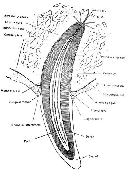
Photo 1A: Normal tooth, support and anatomy.
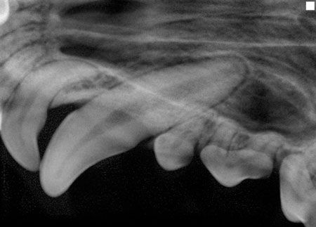
Photo 1B: Normal left maxillary canine, incisor and premolar radiograph. Note the periodontal ligament space surrounding the tooth roots.
Determining the appropriate therapy depends on several factors: the severity of damage to the tooth structure, the periapical and periodontal pathology, the functional significance of the tooth, available equipment, and your ability to either perform the therapy or refer the case. When presented with a complicated crown fracture with exposed pulp or a discolored crown secondary to pulpitis, you have two choices: either extract the tooth or perform endodontic therapy, which usually allows the tooth to be saved and returned to function.
Intraoral imaging provides you with the information you need to formulate a treatment plan to best manage endodontic pathology. You can examine the dental film for both internal and external (periapical) endodontic abnormalities, as described below.
Internal abnormalities
Pulpitis caused by thermal or concussive trauma often results in a discolored tooth, an injured pulp and, in time, a nonvital tooth. Treatment is extraction or root canal therapy.
Once the pulp becomes nonvital, it ceases to produce dentin. Radiographic evidence of pulpal death varies, from an enlarged root canal when compared with the adjacent teeth to no change at all (Photos 2A and 2B).
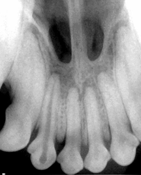
Photo 2A: Discolored right maxillary second incisor revealing enlarged root canal compared with the adjacent teeth.
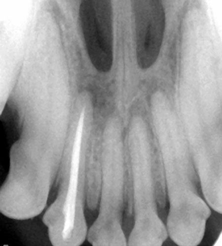
Photo 2B: Root canal therapy performed to save the tooth.
Internal resorption arises from the pulp. The cause is unknown, but trauma or pulpal death from anachoresis (bacteria gaining access to the injured pulp through vascular channels) are believed to be contributing factors. Radiographically the canal will appear irregular or segmentally enlarged (Photos 3A and 3B).
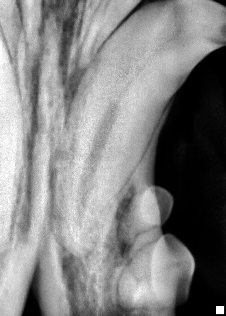
Photo 3A: Internal resorption affecting a left mandibular canine. Note the variance in the root canal diameter.
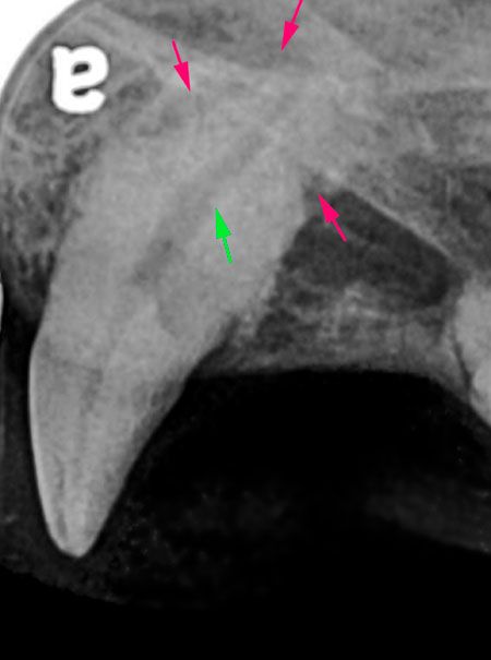
Photo 3B: Internal resorption (green arrow) and external resorption (red arrows).
External abnormalities
Periapical disease surrounds the apex of one or more roots caused by either inflammation or necrosis of the dental pulp from trauma, infection or as an extension of periodontal disease.
The radiographic appearance consistent with a periapical granuloma is a widening of the apical periodontal ligament space with circumscribed alveolar bone resorption (Photo 4). In the early stages of the inflammatory response, minimal bony changes are radiographically evident (Photos 5A and 5B). A periapical cyst, which usually arises from a preexisting granuloma, appears as a sharply outlined circumscribed radiolucent area (Photo 6). Condensing osteitis and osteosclerosis occurs in response to low-grade infection. They appear as radiodense areas around the apex of a vital or nonvital tooth.
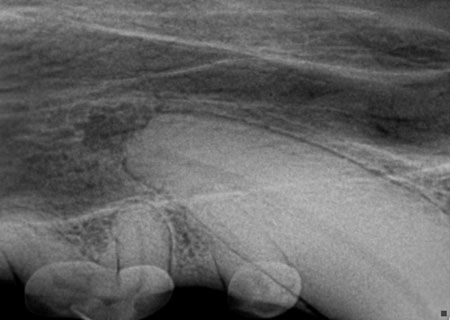
Photo 4: Radiographic appearance of periapical granuloma.
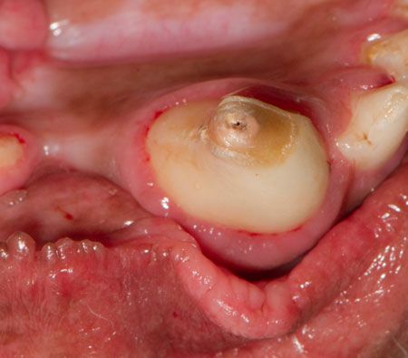
Photo 5A: Complicated mandibular canine crown fracture.
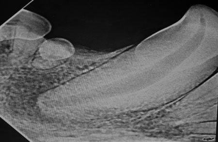
Photo 5B: Early pulpal infection extending periapically.
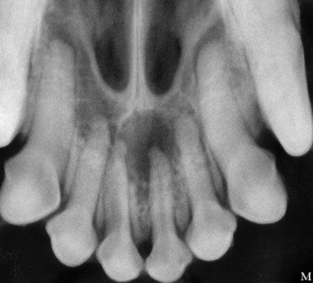
Photo 6: Periapical cyst.
Extraction or root canal therapy are the treatment options if it is determined the tooth is nonvital (Photo 7). A radiolucent artifact (the chevron effect) is a normal outline of the periodontal ligament at the apex, which is sometimes mistaken for pathology (Photo 8).
External root resorption may result from periapical inflammation, excessive occlusal forces or from unknown stimuli. Radiographically, external resorption will appear as radiolucent defects, which can occur on any area of the root surface. Generally the pulp is not affected. When the external resorption extends to the oral cavity, extraction is indicated (Photo 9).
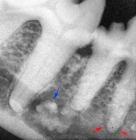
Photo 7: Condensing osteitis (blue arrow) and periapical lucency consistent with pulpal necrosis (red arrows).
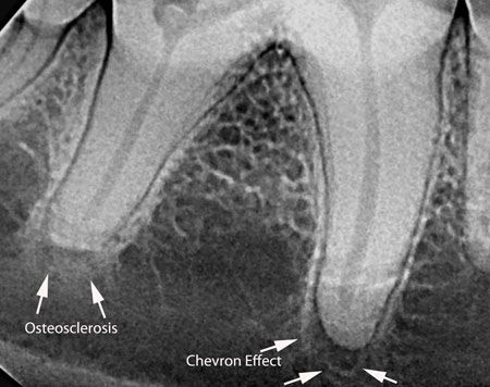
Photo 8: Osteosclerosis and chevron effect.
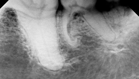
Photo 9: External root resorption of the left mandibular second molar mesial root.
Endodontic and periodontal lesions around the same tooth
Class 1 periodontal-endodontic lesions are primary endodontic lesions that extend toward the crown from the root apex, eventually reaching the gingival sulcus, causing a secondary periodontal lesion. The pattern of bone loss often resembles a J shape (Photo 10).
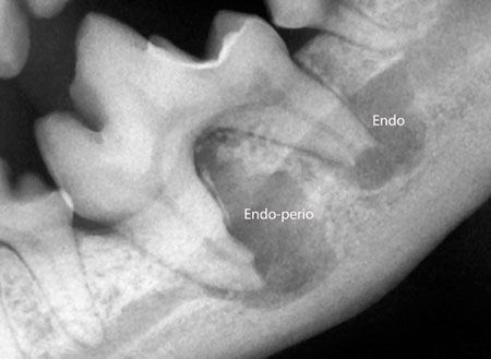
Photo 10: Primary endodontic lesion with secondary periodontal involvement.
Class 2 periodontal-endodontic lesions occur when the loss of attachment extends toward the root apex to a lateral canal or to the apical delta leading to pulpal necrosis. Class 2 periodontal-endodontic lesions often affect the mandibular first molar and appear as one “floating” root without alveolar support, with a periapical endodontic lesion affecting the other tooth root (Photo 11).
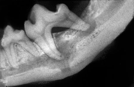
Photo 11: Primary periodontal disease and secondary endodontic periapical radiolucency.
Class 3 combined periodontal-endodontic lesions are true combined, but separate endodontic and periodontal lesions which have coalesced (Photo 12).
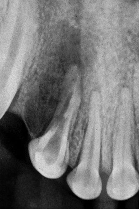
Photo 12: Coalesced endodontic and periodontal lesions in a maxillary second incisor.
When confronted with a patient with a fractured or discolored tooth, intraoral dental radiography paves the road for more thorough evaluation and the development of an appropriate treatment plan. For more information, head over to avdc.org.
Dr. Jan Bellows owns Hometown Animal Hospital and Dental Clinic in Weston, Fla. He is a diplomate of the American Veterinary Dental College and the American Board of Veterinary Practitioners. He can be reached at (954) 349-5800; e-mail: [email protected]