Using thermal imaging in feline care
John C Godbold Jr, DVM, explained the colors that show with temperature-sensing technology and how they can lead to a diagnosis
When looking at a thermal image, practitioners can observe different colors located all over their patients. Reds, blues, yellows, and other colors light up the image to paint a picture of what is happening within patients.
Presenter John C. Godbold Jr, DVM, Stonehaven Veterinary Consulting in Jackson, Tennessee, explained how thermal imaging can help treat cats, and lead practitioners to a diagnosis of their patient in a noninvasive, noncontact, and nonthreatening way, during his lecture at the 2022 American Association of Feline Practitioners conference in Pittsburgh, Pennsylvania.
Thermal imaging 101
Using thermal imaging, veterinary practitioners will notice that when scanning a patient with normal blood flow, the colors will be symmetrical within a patient. The temperatures within the image will present as nonsymmetrical in a patient with abnormal blood flow or suffering from a disease.
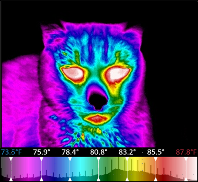
“Now, let me interject when we see thermal images, almost all of them I'm going to show you colors that represent the temperatures. The colors in the palette are very intuitive, ranging from what we mentally think of as being cold, which is black and purple, ranging up from blue, green, yellow, orange, red, and white as the hottest temperatures. But we've got nonsymmetrical temperature distribution here, and there has to be a physiological reason for that,” said Godbold.
Thermal image of feline (Courtesy of Godbold)

He explained that color changes can be caused by an increase in temperature because of hyperthermia and increased blood flow from an inflammation or infection. Practitioners may see a decrease in blood flow because of neurological damage, vasoconstriction, or infarction.
Godbold informed attendees that thermal imaging provides a different way to see what is happening within a patient besides the normal scans because it gives a different point of view and provides a more comforting approach for their feline patients.
“Keep in mind as we talk about this, that traditional imaging that we've done for years with a radiograph, CT scans, MRIs, and ultrasounds give us anatomical information, they show us structurally what's going on. What’s really different about thermal images is we're getting functional and physiological information. The thermal images tell us what's going on in the tissue right now and tell us where we've got warm areas [and] where we've got cooler areas in relation to each other,” said Godbold.
“Now, the beauty of thermal imaging is that this is part of what makes it so cat friendly, is it's noninvasive, we're not sending anything to the cat, we're simply measuring the temperatures on the surf body surface area of the cat. It's absolutely objective. It's just a collection of data, and it is incredibly quantitative data,” he added.
Seeing the unseen
Thermal imaging can be used for wellness screens, patients that are sick, and geriatric patients. Godbold informed attendees that these images are not diagnostic, but they can help provide early detection of physical problems often before any structural changes show. This can be a crucial step for practitioners because if the images show a thyroid issue or issues with a patient's legs, potentially gives them an early diagnosis, early treatment, and possibly a better outcome for both patients, and pet owners.
“I think of thermal damages as being part of the breadcrumb trail that we can use when we're working with patients. They let us be proactive rather than reactive because thermal images detect problems so early in the process of disease. So, we can identify areas requiring further evaluation and these thermal images, and we'll talk about this case in a little more detail later,” said Godbold.
When looking at the images, some parts of the body will present hot, and others will typically always present as cold. On the hot side, the eyes and the anus are usually red because they are consistently warmer, while a cat's tail will typically be present with colder colors.
Conclusion
As medical professionals, Godbold understands that evidence behind technology, like this one, is an important part of implementing it in clinics. If the product has no scientific evidence to back up its significance, there will not be a push to use it within clinics. According to Godbold, the evidence is overwhelming from previous technology he worked with before it entered the veterinary realm.
“I've had the opportunity to work with multiple technologies in this whole realm with lasers and light-based modalities over the years. As they enter veterinary medicine, like everybody else, I want to see the evidence and I want to see veterinary-specific evidence. I can honestly say there is more evidence, veterinary-specific and species-specific, for using thermal imaging than any other technology I have worked with,” he said.
Although successful in studies, according to Godbold, thermal imaging cannot be a stand-alone product, and he warned attendees to be aware of its limits when using the product. It does not provide a specific issue as other imaging can, but it can help by telling veterinary professionals where to start looking if they suspect any issue.
In concluding his lecture, he left attendees with this final thought. “A little marketing slogan that was used for years with this terminology was ‘see the pain.’ and we don't see pain with thermal images. We see physiological changes, and we see alterations in blood flow, but can thermal images help us identify areas that we need to check more closely to see if pain is present? Absolutely,” he concluded.
Reference
Godbold J. Feline Thermal Imaging: What the Pretty Colors Tell Us. Presented at: 2022 American Association of Feline Practitioners Conference, Pittsburgh, Pennsylvania. October 27-30, 2022.