Veterinary leech application to treat toe swelling in a cat
See how medicinal leeching helped this cat get back in its feet.
A 1-year-old castrated male domestic shorthaired cat was presented to a veterinary clinic for evaluation of acute nonweightbearing left hindlimb lameness from an unknown trauma. The cat was an indoor-outdoor pet with no other health problems reported by the owner and was receiving no medications.
On physical examination, no abnormalities were noted except for left pelvic limb crepitus and swelling over the distal tibia. Radiographs of the limb showed a closed, long, oblique, mid-diaphyseal tibial and fibular fracture. The limb was placed in a bivalve cast, and the patient was sent home with pain medication.
At a cast change about two weeks after the placement of the cast, irritation was noted on the lateral aspect of the tarsus where the tape stirrup was placed. The full cast was replaced, and the owner was told to bring the patient back in two weeks.
Four days later, the owner noticed blood and swelling of the toes and brought the patient in for an evaluation. The patient was sedated, and the cast was removed. There was now medial and lateral skin irritation under the tape stirrups, and all of the digits were swollen, erythematous and oozing a serosanguinous fluid through the skin. No obvious skin wounds were present, so the cast was replaced. The fracture was palpated, and a callus was present.
The next day the patient returned because of swelling of the toes and pain on ambulation. The cast and padding were removed, and partial-thickness abrasions were noted with severe swelling of the digits. The owners reported that the patient had not been using the limb since the third cast change and now was not even getting up. On physical examination, the patient was febrile (104 F [40 C]), growling and exhibiting pain on palpation of the limb. The patient refused to ambulate but had motor function in all four limbs. The entire paw was so swollen that the nail beds were not visible.

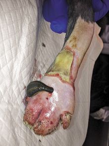
Figure 1. Two leeches were applied to the dorsal surface of the cat's paw. They fed until they detached spontaneously. Photos courtesy of Dr. Nicole J. Buote.There was moderate serosanguinous discharge within the cast padding and partial-thickness wounds along the dorsal and ventral paw and white discolored skin laterally and medially with clear demarcation of healthy vs. unhealthy tissue. There was normal sensation to the paw. The paw was clipped and cleaned with chlorhexidine and sterile saline solution, the partial-thickness wounds were gently débrided and a soft padded bandage with a splint was placed. Twice-daily bandage changes were done the next day, and the patient was discharged with the same antibiotics and pain medication as before.
The cat underwent bandage changes every two days for the next six days. On the sixth day, the wound was found to be a full-thickness, 360-degree skin wound of the distal metatarsal tarsal region with severe swelling of the toes, most likely from constriction from the bandage. The paw still had good motor function and sensation, and the tibial fracture site palpated stable with moderate callus. Because the paw swelling had not resolved even after débridement of the constrictive wound and compressive bandages, the differential diagnosis at this time was venous stasis from the constrictive wound rather than interstitial edema.

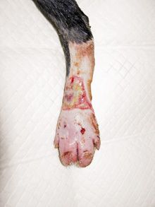
Figure 2. Leech application was performed daily on the cat for the next four days, and the patient's paw size reduced dramatically.A decision was made to try therapeutic leeching because treatments for interstitial edema had failed, and leeching is a well-known treatment for venous stasis in people. The mechanism behind the leeching process and potential complications were explained to the owners, and they consented.

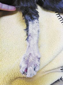
Figure 3. Nine weeks after the initial injury, the cat had full use of its limb. No complications from the leeching procedure were identified.The patient was sedated with dexmedetomidine and buprenorphine. Any remaining questionable tissue was débrided, and the paw was cleaned and dried. Two leeches were applied to the dorsal surface of the paw (Figure 1), and they fed until they detached spontaneously. A temporary soft-padded bandage was applied to absorb residual oozing, and then a new soft-padded bandage with a splint was applied two hours later. Leech application was performed daily for the next four days, and the patient's paw size reduced dramatically (Figure 2). The patient's limb became weight-bearing after two days of the leeching treatment.
The patient received antibiotics (13.75 mg/kg amoxicillin-clavulanic acid orally b.i.d.) for two weeks. With the leech treatment, bandage changes continued every two to seven days for another two weeks until the wound was healed. The splint was then removed, and a soft-padded bandage was applied for another two weeks until radiography showed adequate healing of the tibial and fibular fracture (nine weeks after the original injury). At that time, the patient had full use of its limb, and no complications from the leeching procedure were identified (Figure 3).