Adverse vaccine reactions in veterinary medicine: an update
Although rare, several forms of adverse reactions have been documented after vaccination in dogs and cats. Heres what veterinary teams need to know today.

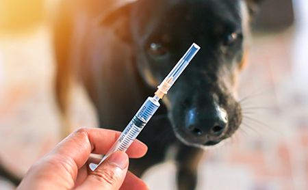
Trsakaoe/stock.adobe.comWhile approved vaccination protocols are considered safe for the vast majority of canine and feline patients, adverse vaccine reactions can occur in veterinary medicine. Many cases are associated with either misuse of the vaccine or overvaccination.
An updated overview of the various forms of adverse vaccine reaction was provided last year in Veterinary Clinics of North America: Small Animal Practice.1 The author, Laurel J. Gershwin, DVM, PhD, DACVM, a long-time immunology professor at the University of California, Davis, serves on the American Veterinary Medical Association (AVMA) Council on Biologic and Therapeutic Agents, which oversees vaccination principles for the association. In her article, Dr. Gershwin described the current understanding of adverse vaccine reactions in veterinary medicine, including type I and type III hypersensitivities, vaccine-associated fibrosarcoma, vaccine-associated disease enhancement and formation of autoimmune disease.
Type I hypersensitivity
A type I hypersensitivity vaccine reaction, consisting of an immunoglobulin E (IgE) response to nontarget proteins, most commonly occurs after administration of inactivated, adjuvanted viral vaccines. Nontarget proteins are not associated with the virus itself, but rather originate either in the growth medium used to amplify the virus (e.g. fetal bovine serum or egg protein) or from the addition of stabilizers (including gelatin and casein) to aid storage of the vaccine. While it is not uncommon for a small amount of IgG production to occur against these nontarget proteins, animals that are prone to hypersensitivity-associated diseases, including horses and dogs, are more likely to stimulate inappropriate IgE production.
IgE antibodies function by binding to Fc receptors on basophils as well as mast cells located in the skin and mucous membranes of the respiratory and intestinal tracts. When an adverse vaccine reaction occurs, nontarget antigen in the vaccine binds to IgE, resulting in mast cell degranulation, histamine release and activation of the lipoxygenase and cyclooxygenase pathways. In dogs, type I hypersensitivity results in increased capillary permeability and smooth muscle contraction, causing swelling and urticaria of the muzzle and, in severe cases, anaphylaxis. Horses experiencing type I hypersensitivity after vaccination may display colic or anaphylactic shock. Hypersensitivity usually repeats with subsequent vaccinations, as different vaccines often contain the same nontarget antigens.
In one experimental population of beagles, administration of a rabies vaccine containing alum adjuvant resulted in increased IgE production against not only vaccine antigens but also bovine serum proteins.2 Results from another study involving a population of Maltese-beagle crossbred dogs known to be allergic to corn and soy showed that administration of core vaccines caused a significant increase in IgE production against the food allergens.3 This study's results implied that vaccination may worsen food allergies in susceptible individuals. Finally, a third study found that dogs that reacted adversely to vaccines had relatively high amounts of circulating IgE against vaccine adjuvants and stabilizers compared with nonreactive dogs.4 A similar study determined that horses that were highly reactive to vaccination also reacted to adjuvant on intradermal testing.5
Dr. Gershwin suggested two alternatives to vaccination for small animal patients that are suspected to react adversely to the rabies vaccine, a required immunization in all 50 states. Some states allow serum antibody titer measurement to substitute regular administrations of the rabies vaccination. Alternatively, 0.1 mL of the vaccine can be administered as an intradermal skin test, using histamine as a positive control and either vaccine diluent or sterile saline as a negative control. If a wheal develops after 15 to 20 minutes, then the patient is generating an IgE response and is likely to experience an adverse reaction upon vaccination.
Type III hypersensitivity
The Arthus reaction, or type III hypersensitivity response, is characterized by formation of antigen-antibody immune complexes. Specific IgG circulating at the time of vaccination binds to target or nontarget protein antigens, leading to mast cell degranulation, neutrophil chemotaxis, and subsequent local hemorrhage and inflammation.
Arthus reaction occurs locally at the injection site within 24 hours of vaccination and, while the site may be swollen and painful, the response is usually not serious and resolves within 2 to 3 days. Dr. Gershwin recommended measuring serum antibody titer in patients that experience type III hypersensitivity before giving additional doses of the suspect vaccine.
Vaccine-associated fibrosarcoma
Sometimes referred to as injection-site sarcoma, vaccine-associated fibrosarcoma was first reported in cats in 1991.6,7 This adverse reaction is rare, with an estimated occurrence in cats of 0.01% to 0.1%. It is most commonly associated with administration of adjuvanted, killed vaccines such as rabies and feline leukemia virus (FeLV). The incidence of vaccine-associated fibrosarcoma has apparently not decreased significantly despite the recent addition of a nonadjuvanted rabies vaccine to the market.8
Vaccine-induced disease enhancement
In rare cases, vaccine administration can induce a pathogenic immune response. In veterinary medicine, this effect has occurred in calves vaccinated against bovine respiratory syncytial virus (BRSV), with research showing that the killed form of the vaccine stimulates a T-helper type 2 response involving IgE antibody production rather than the more appropriate T-helper type 1 response necessary for viral clearance.9 This effect appears to be dose dependent.10
Vaccine-induced enhancement has also been reported after administration of the feline infectious peritonitis (FIP) vaccine. With this vaccine, researchers observed that IgG antibodies produced after vaccination actually activated macrophages, thus facilitating the occurrence of antibody-dependent disease enhancement that is necessary for coronavirus to spread through the body. The FIP vaccine is currently listed as “not generally recommended” by the American Association of Feline Practitioners (AAFP).11
Autoimmune disease development
Dr. Gershwin described “scant” evidence for the development of autoimmune diseases, such as immune-mediated hemolytic anemia (IMHA) and hypothyroidism, as a result of vaccination. One study found that 26% of examined dogs with IMHA developed the disease within 1 month after receiving one or more common vaccines, including canine distemper virus, parvovirus, leptospirosis bacterin, and Bordetella bronchiseptica bacterin.12 However, other studies examining similar factors were unable to confirm any association between IMHA and vaccination.13
A study determined that dogs that had received a multivalent vaccine that included a killed, alum-adjuvanted rabies vaccine had significantly higher circulating antibody to antithyroglobulin than unvaccinated dogs did.14 However, dogs that received another form of multivalent vaccine lacking the rabies component did not have high antibody levels. The authors of that study proposed that thyroglobulin contained in the fetal bovine serum used to grow the virus may have stimulated the immune reaction.
Despite little evidence for an association between vaccination and autoimmune disease, it is undeniable that many dog breeds are predisposed to certain autoimmune diseases-such as Samoyeds-and diabetes mellitus. Detailed research found that differences in canine histocompatibility antigen haplotypes likely account not only for diabetes in Samoyeds but for genetic predispositions of other breeds to other autoimmune diseases as well.15 Therefore, Dr. Gershwin emphasized that the causes of autoimmune disease are likely multifactorial and include genetics and environmental factors as well as immune dysfunction.
Recent changes in vaccination guidelines
Many professional veterinary organizations have instituted changes to vaccination guidelines to further minimize risk of adverse vaccine reaction development. The Vaccine-Associated Feline Sarcoma Task Force (VAFSTF) and AAFP now indicate that core feline vaccines should be administered in specific anatomic locations. The rabies vaccine is to be administered as distally as possible on the right rear limb and the FeLV vaccine on the distal left rear limb. All other vaccines should be administered to the right shoulder, but not the midline or intrascapular space. In the event of fibrosarcoma formation, the suspected vaccine can be identified and the affected limb can be amputated if these guidelines are followed.
Also, Dr. Gershwin cited extensive evidence that most canine and feline core vaccines have a greater duration of effectiveness than previously believed and likely provide at least 3 years of coverage. Therefore, the AVMA, American Animal Hospital Association, and AAFP recommend booster vaccinations to be administered every 3 years for dogs and cats following the puppy/kitten series and the initial 1-year booster. This change, Dr. Gershwin noted, will further reduce the risk and incidence of adverse reaction after vaccination.
References
Gershwin LJ. Adverse reactions to vaccination: from anaphylaxis to autoimmunity. Vet Clin Small Anim 2008;48:279-290.
HogenEsch H, Dunham AD, Scott-Moncrieff C, et al. Effect of vaccination on serum concentrations of total and antigen specific immunoglobulin E in dogs. Am J Vet Res 2002;63(4):611-616.
Tater KC, Jackson HA, Paps J, et al. Effects of routine prophylactic vaccination or administration of aluminum adjuvant alone on allergen-specific serum IgE and IgG responses in allergic dogs. Am J Vet Res 2005;66(9):1572-1577.
Ohmori K, Masuda K, Maeda S, et al. IgE reactivity to vaccine components in dogs that developed immediate-type allergic reactions after vaccination. Vet Immunol Immunopathol 2005;104(3-4):249-256.
Gershwin LJ, Netherwood KA, Norris MS, et al. Equine IgE responses to non-viral vaccine components. Vaccine 2012;30(52):7615-7620.
Kass PH, Barnes WG Jr, Spangler WL, et al. Epidemiologic evidence for a causal relation between vaccination and fibrosarcoma tumorigenesis in cats. J Am Vet Med Assoc 1993;203:396-405.
Hendrick MJ, Brooks JJ. Postvaccinal sarcomas in the cat: histology and immunohistochemistry. Vet Pathol 1994;31:126-129.
Wilcock B, Wilcock A, Bottoms K. Feline postvaccinal sarcoma: 20 years later. Can Vet J 2012;53(4):430-434.
Gershwin LJ, Schelegle ES, Gunther RA, et al. A bovine model of vaccine enhanced respiratory syncytial virus pathophysiology. Vaccine 1998;16(11-12):1225-1236.
Schneider-Ohrum K, Cayatte C, Bennett AS, et al. Immunization with low doses of recombinant postfusion or prefusion respiratory syncytial virus F primes for vaccine-enhanced disease in the cotton rat model independently of the presence of a Th1-biasing (GLA-SE) or Th2-biasing (Alum) adjuvant. J Virol 2017;91(8):e02180-16.
Scherk MA, Ford RB, Gaskell RM, et al. 2013 AAFP feline vaccination advisory panel report. J Feline Med Surg 2013;15(9):785-808.
Duval D, Giger U. Vaccine-associated immune-mediated hemolytic anemia in the dog. J Vet Intern Med 1996;10(5):290-295.
Carr AP, Panciera DL, Kidd L. Prognostic factors for mortality and thromboembolism in canine immune-mediated hemolytic anemia: a retrospective study of 72 dogs. J Vet Intern Med. 2002;16(5):504-509.
Scott-Moncrieff JC, Azcona-Olivera J, Glickman NW, et al. Evaluation of antithyroglobulin antibodies after routine vaccination in pet and research dogs. J Am Vet Med Assoc 2002;221(4):515-521.
Kennedy LJ, Davison LJ, Barnes A, et al. Identification of susceptibility and protective major histocompatibility complex haplotypes in canine diabetes mellitus. Tissue Antigens 2006;68(6):467-476.
Dr. Natalie Stilwell provides freelance medical writing and aquatic veterinary consulting services through her business, Seastar Communications and Consulting. In addition to her DVM obtained from Auburn University, she holds a MS in fisheries and aquatic sciences and a PhD in veterinary medical sciences from the University of Florida.
