Busting oncology myths
Experts debunk common misconceptions in veterinary oncology
Myth 1: Most masses in patients younger than 12 months of age are nonneoplastic
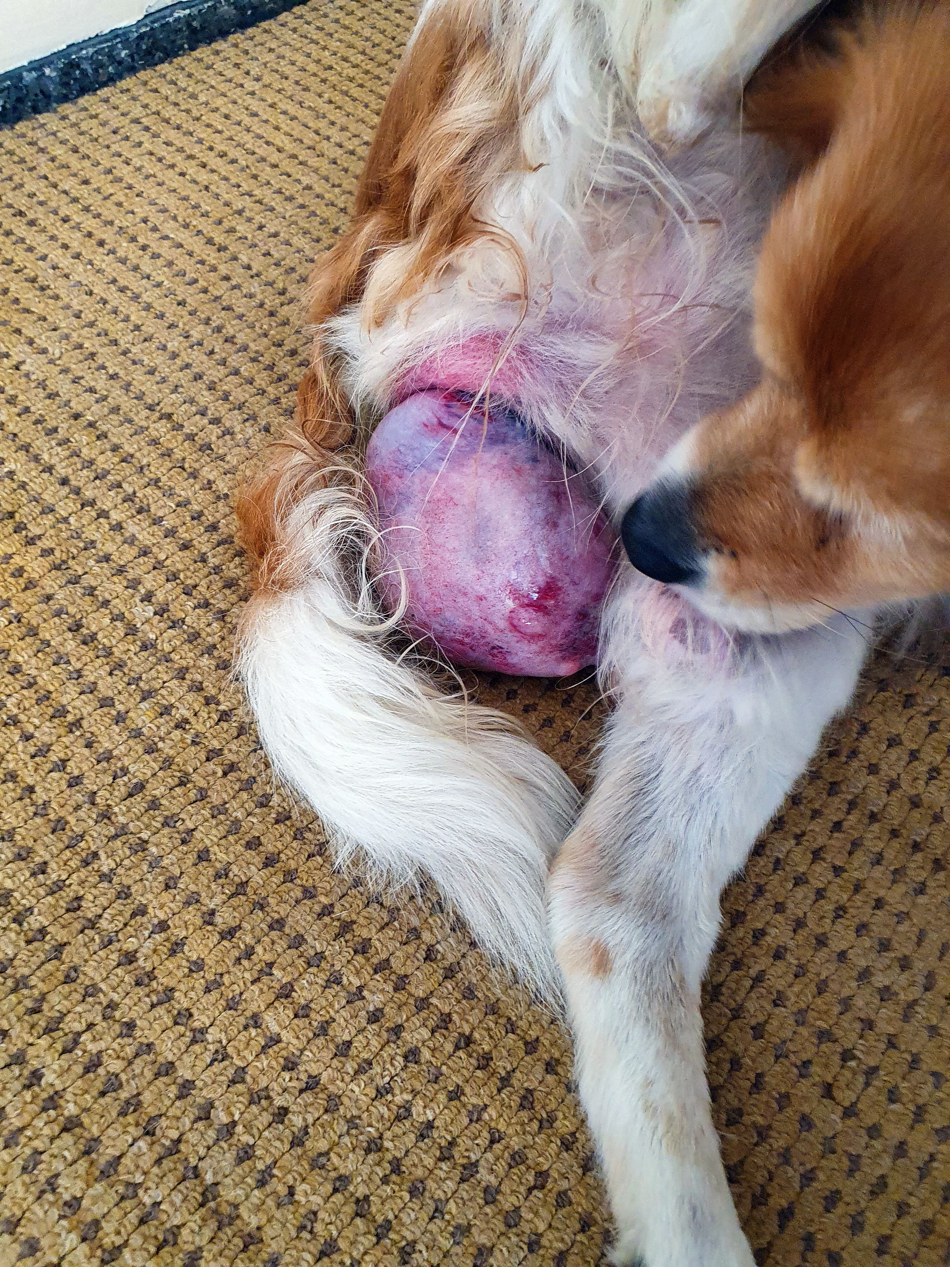
This is considered semantics to some veterinary practitioners and academic to others, but that approach is technically very inaccurate. In a recent study by Kim et al, 2554 histopathologic samples were submitted from dogs younger than 12 months of age. In total, 2408 of 2554 (94.3%) were neoplastic. Of those, 2212 of 2408 (86.6%) were considered benign neoplastic and were interpreted as histiocytoma.1 Veterinarians often do not understand and/or document the difference between benign masses (eg, cyst, viral, hyperplasia, granuloma), benign neoplastic masses, and malignant neoplastic masses and/or appropriately communicate this information to pet owners.
Neoplasia simply means new, abnormal growth; it does not indicate a benign or malignant process. For a cell to become neoplastic, it develops mutations that allow it to no longer obey that boundary of local cells, thus allowing uncontrolled growth. To classify as a benign neoplastic mass, the abnormal cells stay in 1 place vs a malignant neoplasm, where the cells can metastasize. Although many histiocytic tumors can spontaneously regress and not need surgery, the location and progression of growth must be carefully monitored to not lose a window where surgical removal is less complicated.
Myth 2: Canine cutaneous and subcutaneous mast cell tumors have the same grading system and surgical recommendations
Pathologists use the Kiupel grading system (low vs high grade) and/or the Patnaik grading system (I, II, III) for cutaneous mast cell tumors (MCTs). Patnaik grade I and most grade II MCTs are considered low grade per the Kiupel grading system. Patnaik grade III is considered high grade in the Kiupel grading system. The grade is associated with metastatic potential.
As reported by Simpson et al in 2004, 2-cm margins lateral and 1 fascia plane deep are recommended for cutaneous MCTs. In this study, 100% of grade I MCTs were completely excised at 1-cm lateral margins and 1 fascia plane deep. Seventy-five percent of grade II MCTs were excised at 1-cm lateral margin and 1 fascia plane deep, with 100% excised using the 2-cm margin and 1 fascia plane deep. No grade III MCTs were present in the study population.2
The Kiupel and Patnaik grading systems cannot be applied to MCTs that are confined to the subcutaneous space (SQ). SQ MCTs that are completely restricted to the fat do not have a grading system. Moreover, there have yet to be published studies to document the recommended margins. However, most surgeons still have the goal of achieving as close to 2 cm lateral and 1 fascia plane deep for margins.
In general, most subcutaneous MCTs have a favorable prognosis, with extended survival times (84% alive at 1500 days), low rates of reoccurrence (2%), and low metastatic rates (6%). Thompson et al concluded that SQ MCTs were more effectively controlled by surgery alone (98%) compared with cutaneous MCTs.3,4
Myth 3: Agreement between cytologic findings and histopathologic findings is usually at least 75%
annebel146 / stock.adobe.com

According to Cohen et al in the Journal of the American Veterinary Medical Association in 2003, complete agreement between cytology and histopathologic diagnosis was 37.9% (102 of 269), with complete disagreement in 32.7% (88 of 269) of cases. When insufficient cytologic samples were removed from submission, there was 42.7% complete agreement.5 These results contrast with previously published studies by Eich et al and Vos et al, which had sensitivity of 89%/95.6% and specificity of 100%/65.4%.6,7 The key difference is that the cytologic samples for these reports were obtained during surgery or postmortem and not in awake, moving patients.
Cytology slides should be assessed prior to submission to reduce the chances of rejection because of low cellularity, cell degeneration, hemodilution, and/or artifacts. At least 1 slide should be assessed in-house before submission. In addition, multiple locations within a mass should be aspirated to increase the odds of obtaining an accurate diagnosis.
Tasha Ostrowski, RVT, graduated from Kansas State University in 2017 with a major in animal sciences and industry, then went on to attend Colby Community College and became a registered veterinary technician in 2020. Since then, she has been working as a surgery nurse in a multispecialty practice while awaiting 2023 acceptance letters into veterinary school. Her dream is to become a veterinarian and continue her passion to care for the animals that bring joy into our lives, no matter the size.
Heather Towle Millard, DVM, MS, DACVS-SA, is a staff surgeon at Phoenix Veterinary Surgical Specialists. With over 15 years of experience, Millard’s veterinary expertise includes advanced soft-tissue surgery, oncologic surgery, and general orthopedic surgery. She enjoys mentoring passionate, energetic, and determined young doctors and being part of their journey to success.
References
- Kim D, Dobromylskyj MJ, O’Neill D, Smith KC. Skin masses in dogs under one year of age. J Small Anim Pract. 2022;63(1):10-15. doi:10.1111/jsap.13418
- Simpson AM, Ludwig LL, Newman SJ, Bergman PJ, Hottinger HA, Patnaik AK. Evaluation of surgical margins required for complete excision of cutaneous mast cell tumors in dogs. J Am Vet Med Assoc. 2004;224(2):236-240. doi:10.2460/javma.2004.224.236
- Thompson JJ, Pearl DL, Yager JA, Best SJ, Coomber BL, Foster RA. Canine subcutaneous mast cell tumor: characterization and prognostic indices. Vet Pathol. 2011;48(1):156-168. doi:10.1177/0300985810387446
- Weisse C, Shofer FS, Sorenmo K. Recurrence rates and sites for grade II canine cutaneous mast cell tumors following complete surgical excision. J Am Anim Hosp Assoc. 2002;38(1):71-73. doi:10.5326/0380071
- Cohen M, Bohling MW, Wright JC, Welles EA, Spano JS. Evaluation of sensitivity and specificity of cytologic examination: 269 cases (1999-2000). J Am Vet Med Assoc. 2003;222(7):964-967. doi:10.2460/javma.2003.222.964
- Eich CS, Whitehair JG, Moroff SD, Heeb LA. The accuracy of intraoperative cytopatholog- ical diagnosis compared with conventional histopathological diagnosis. J Am Anim Hosp Assoc. 2000;36(1):16-18. doi:10.5326/15473317-36-1-16
- Vos JH, van den Ingh TS, van Mil FN. Non-exfoliative canine cytology: the value of fine needle aspiration and scraping cytology. Vet Q. 1989;11(4):222-231. doi:10.1080/0165217 6.1989.9694228

Newsletter
From exam room tips to practice management insights, get trusted veterinary news delivered straight to your inbox—subscribe to dvm360.