Carcinomas found with most clinically detected thyroid tumors
Signalment: Canine, Beagle, 6-year-old, male castrated, 62 lbs.
Signalment:
Canine, Beagle, 6-year-old, male castrated, 62 lbs.
Clinical history:
The dog presents for coughing, gagging, some polyphagia and pica, and polydipsia. There has been no history of dysphagia.

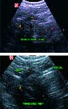
Photo 1 (top) Photo 2 (bottom)
Physical examination:
The findings include rectal temperature 101.3° F, heart rate 120/min, respiratory rate 35/min, pink mucous membranes, normal capillary refill time, body condition score 4/5, and normal heart and lung sounds. There also are mild submandibular lymph node enlargement and a thickening distal to the larynx - suspect involvement of the thyroid gland.
Laboratory results:
A complete blood count, serum chemistry profile and urinalysis are outlined in Table 1.
Thyroid panel:
The serum T4 value (RIA) is 8.79 (normal range 1.0-4.0 µg/dl); the serum free T4 value (equilibrium dialysis) is 89 (normal range 11-43 pmol/l). The dog has not been receiving any thyroid supplementation.
Ultrasound examination:
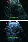
Proximal cervical ultrasonography was performed. The ultrasound images provided are from the thickened area immediately distal to the larynx.

Photo 3 (top) Photo 4 (bottom)
My comments:
There is an echogenic mass with distinct margins present distal to the larynx that could represent a mass involving the thyroid gland.
Case management:
In this case, most likely neoplasia of the thyroid gland is the clinical diagnosis. At this point, I would recommend thoracic radiographs be done to check for metastatic disease in the lungs and cranial mediastinum. Also, fine-needle aspirations of the enlarged submandibular lymph node and echogenic cervical mass for cytologic examination would be recommended.
The treatment for this dog is possible surgical excision of the echogenic mass and confirmation by histopathologic examination as to the cell type of the neoplasia. Thereafter, a chemotherapy protocol may be established because I would suspect metastatic disease to be present.
Thyroid tumor review
Thyroid tumors are relatively common in middle-aged or older dogs; most clinically detected thyroid tumors are carcinomas that locally invade into adjacent structures such as esophagus, trachea, cervical musculature, nerves and thyroid vessels, and generally produce distant metastasis.

Thyroid carcinomas are usually large, easily palpable and result in clinical signs caused by invasion or compression of local tissues that can be recognized by the owner. There is no sex predilection. Breeds reported to have an increased risk of developing thyroid neoplasia include Boxers, Beagles and Golden Retrievers.
Many dogs with thyroid tumors are examined because the owner has noticed an enlargement of the neck. Most thyroid tumors are large, easily palpable and fixed to the soft tissues of the neck. Signs may include dyspnea, cough, hoarseness, dysphagia, vomiting, anorexia and weight loss, especially in dogs with nonfunctional thyroid tumors.
In addition, dogs with nonfunctional thyroid tumors are usually euthyroid.
Hyperthyroidism
Of all thyroid tumors in dogs, approximately 10 percent are autonomous and hyperfunctional, leading to signs of hyperthyroidism. As in dogs with nonfunctional tumors, most hyperthyroid dogs have a thyroid carcinoma.

Table 1
In general, hyperfunctional tumors causing hyperthyroidism are usually smaller than the nonfunctional tumors and have less a compressive effect on adjacent structures. polyuria and polydipsia are usually the earliest and most predominant signs associated with hyperthyroidism in dogs. Weight loss, despite a good appetite, is also common.
Other signs that may develop in dogs with hyperthyroidism include weakness, fatigue, heat intolerance, nervousness or restless behavior, and more frequent defecation with passage of semiformed stools. Cardiac signs may include a more forceful apex beat and arterial pulse; the ECG may show high voltage in all leads.
Ventral cervical region mass
Thyroid neoplasia should be suspected in any dog with an enlarging mass in the ventral cervical region. Other differential considerations would include abscesses, granulomas and nonthyroidal neoplasia. Fine-needle aspiration cytology may be helpful in differentiating a thyroid tumor from an abscess, salivary mucocele or enlarged lymph node, although thyroid masses are typically quite vascular and one may only retrieve blood.
A definitive diagnosis usually requires an excisional biopsy and histopathologic examination. High serum concentrations of total and free T4 would be expected in dogs with hyperfunctional thyroid neoplasia causing hyperthyroidism. Thoracic radiographs should always be reviewed because about one third of these dogs have pulmonary metastasis at the time of diagnosis. Pertechnetate thyroid scans can aid in demonstrating the location of abnormal thyroid tissues.
Because of the highly malignant nature of this disease, treatment of thyroid neoplasia in dogs is usually not curative. most dogs do experience palliative relief from surgical excision of the smaller thyroid masses.
An attempt at surgical removal or debulking of the thyroid tumor should be the first step in managing these cases. Doxorubicin dosed at 30 mg/m2 body surface area intravenously every three to six weeks until total remission of the tumor occurs or adverse reactions to the chemotherapy develop is effective chemotherapy for thyroid carcinoma in dogs.