Clinical Rounds: Thymoma in an 11-year-old chocolate Lab
This dog was referred to an oncologist because of a mass identified by its primary veterinarian. Work through this case with the team at the University of Tennessee to find out what happens once a patient is referred.
The Clinical Rounds team is from the Department of Small Animal Clinical Sciences, College of Veterinary Medicine, The University of Tennessee, Knoxville, Tennessee.
Clinical rounds coordinator: Isabella Pfeiffer, Dr. med. vet., DACVR (radiologic oncology), DECVIM-CA (oncology, radiation oncology)
Medical oncology: Lark Walters, DVM
Internal medicine: Sarah Schmid, DVM
Clinical pathology: Lisa Viesselmann, DVM
Surgery: Christian Latimer, DVM
Anatomic pathology: Nathan Helgert, DVM
Radiology: Adrien Hespel, DVM, MS, DACVR
Radiation oncology: Isabella Pfeiffer, DMV, DECVIM-CA (oncology, radiation oncology), DACVR (radiation oncology)
Thymomas are an uncommon neoplasm of thymic epithelial cells. They tend to occur in older dogs and are the second-most-common tumor of the cranial mediastinum. Clinical signs are typically related to a space-occupying mass of the thorax and can include lethargy, coughing and dyspnea.

Dr. Lark WaltersParaneoplastic syndromes are common and are reported in up to 67% of dogs with thymoma. Myasthenia gravis is the most commonly reported paraneoplastic syndrome; however, exfoliative dermatitis, hypercalcemia and lymphocytosis are also reported.1 Megaesophagus and recurrent episodes of aspiration pneumonia are frequent complications and are reported in as many as 40% of dogs with this tumor.1
Case presentation
An 11-year-old spayed female chocolate Labrador retriever was referred to the University of Tennessee (UT) oncology service for evaluation of a cranial mediastinal mass. The referring veterinarian originally noted the mass two months before by using thoracic radiography. A complete blood count (CBC) at that time was unremarkable. The patient had been gradually slowing down over the last year but had become worse in the two months prior to presentation at UT. The owners reported episodes occurring every few weeks in which the patient's four limbs would shake and the patient would collapse.
At presentation, physical examination results showed a slightly ataxic gait and weakness, especially in the patient's rear limbs. Muffled heart sounds were also noted. The remainder of the examination was unremarkable.
Tumor staging and treatment
A CBC showed a mature neutrophilia (11,960; range = 2,650-9,800) and mild monocytosis (960; range = 165-850) and eosinophilia (890; range = 0-850). Serum chemistry profile, coagulation assay and urinalysis results were unremarkable. The patient's acetylcholine receptor antibody titer was elevated (7.72 nmol/L; range = < 0.6 nmol/L). Abdominal imaging was unremarkable with the exception of bilateral mildly enlarged adrenal glands. A computed tomography (CT) scan of the patient's thorax was performed.
The mediastinal mass was sampled by using ultrasound-guided fine-needle aspiration. Samples were submitted for cytology and flow cytometry. The results were consistent with thymoma.
The patient was administered pyridostigmine, and its systemic myasthenia gravis signs improved. The patient had no signs of a megaesophagus or esophageal dysfunction at that time. Based on the CT scan results, the tumor was deemed unresectable due to intimate association with the vena cava.
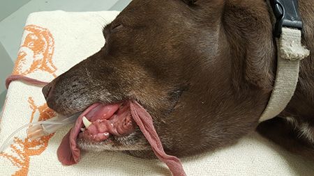
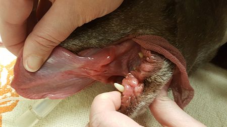
The patient started definitive radiation therapy about three weeks after its initial presentation at UT. At this time, the dog developed edema in its cervical region, most likely due to cranial vena cava compression (Figures 1 & 2). The patient was scheduled to receive 20 fractions on a Monday-to-Friday schedule.

Figure 1. The patient intubated and anesthetized, showing edema of the ventral neck due to compression of the vena cava. Photos courtesy of University of Tennessee.

Figure 2. The patient developed sublingual edema due to caval compression.Follow-up
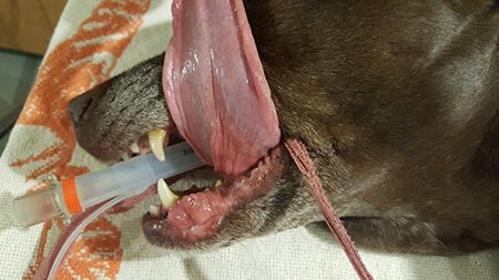
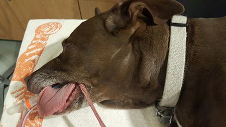
Thoracic radiography was performed 11 days later as half of the patient's radiation therapy protocol was complete. The tumor seemed smaller and more translucent. The dog still had no sign of megaesophagus and was doing well with the treatments. The patient's cervical edema had significantly improved (Figures 3 & 4). Prednisone therapy (0.58 mg/kg/day) was started.

Figure 3. The patient's ventral neck edema resolved after radiation therapy.

Figure 4. The patient's sublingual edema resolved after radiation therapy.Three days later, when the patient was presented to continue radiation therapy, the dog was in severe respiratory distress. Owners reported that the dog had regurgitated multiple times that morning and the day before. Nasal oxygen was started, and thoracic radiography was repeated. Megaesophagus and aspiration pneumonia were diagnosed.
The patient was transferred to the internal medicine service and was treated with intravenous fluids, nasal oxygen, antibiotics (Unasyn-Pfizer), enrofloxacin, maropitant (Cerenia-Zoetis), and nebulization and coupage. Prednisone and pyridostigmine were continued. The patient improved rapidly and was discharged two days later because of financial restrictions.
At discharge, the patient was receiving oral medications, and the owners had a plan to manage the megaesophagus at home and with the referring veterinarian. Radiation therapy was discontinued because of the high risk of aspiration during recovery and because acute adverse effects such as esophagitis may exacerbate esophageal dysfunction.
The patient did well with elevated feedings and oral medications until aspiration pneumonia was diagnosed three months later. Hospitalization was declined, and the patient did not respond to oral medications. The patient was euthanized because of poor clinical condition.
Reference
1. Withrow S, Vail D, Page R. Myeloma-related disorders. In: Vail D, ed. Withrow and MacEwen's small animal clinical oncology. 5th ed. St. Louis: Elsevier, 2013.
Click through the links below to read the different perspectives on this case.