Diagnosing and treating upper airway disease (Proceedings)
Upper airway disorders in dogs and cats include abnormalities of the nares, pharynx, larynx and trachea. The purpose of this manuscript is to highlight the most common upper airway disorders that we seen in canine and feline patients. A more detailed discussion will occur during the lecture.
Upper airway disorders in dogs and cats include abnormalities of the nares, pharynx, larynx and trachea. The purpose of this manuscript is to highlight the most common upper airway disorders that we seen in canine and feline patients. A more detailed discussion will occur during the lecture.
Brachycephalic airway syndrome
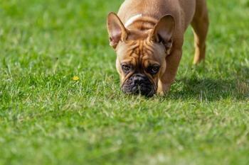
Dogs and cats with brachycephalic airway syndrome typically have some combination of stenotic nares, elongated and thickened small palate, eversion of laryngeal saccules and hypoplastic trachea (dogs). Interestingly, the single biggest obstruction to breathing in dogs with this congenital malformation is the tongue. The tongues of these dogs are not inherently long or thick, but because the skull of these patients is shortened the tongue is relatively too long and too thick for their pharynx.
Stenotic nares is an unhealthy defect and not compatible with excellent quality of life. Dogs with stenotic nares can generate enormous negative intra-pharyngeal pressure during forced inspiration. Over time this causes the soft palate to thicken (due to turbulent air flow-induced edema), and elongate due to the resulting vacuum effect that is created. The negative intrapharyngeal pressure also causes eversion of the laryngeal saccules, further worsening the airway obstruction.
Patients with stenotic nares are candidates for surgical correction of the narrowed opening to their nares; this should be considered at time of spay or neuter or at any time in their life when anesthesia is required (dentistry, laceration repair etc). This is a simple procedure that can be done by any experienced general practitioner and can be successfully accomplished in 15 minutes. Correction of elongated soft palate can be accomplished with traditional surgical techniques or laser or coblation treatment. Because the soft palate can swell as a post operative complication, and because soft palate swelling can result in a life threatening airway obstruction, only experienced surgeons should attempt to shorten an elongated soft palate.
Nasopharyngeal polyps
Nasopharyngeal polyps typically occur in cats less than 1 year of age. The etiology is not understood, although the polypoid tissue generally arises from the lining of the eustachian tube and grows into the area of least resistance including the ear canal and posterior (caudal) choanae. Cats with polyps in this region often present to the veterinarian with noisy breathing that is most obvious during inspiration. Interestingly, these polyps are often much larger than the client (or veterinarian) imagines and can grow to 3-4 cm in length in a 3-4kg feline patient.
The diagnosis may be suggested by the presence of noisy inspiratory breathing in a young cat without nasal discharge. The formal diagnosis requires visualization that may only be possible when the patient is anesthetized. There are two typical surgical approaches to this problem. Bulla osteotomy is a surgical approach that is most likely to offer a permanent cure because during the osteotomy procedure the origin of the polypoid tissue can be visualized and surgically removed. This is a relatively complicated and delicate procedure. In contrast, a polyp lying over the soft palate that is visualized through the mouth can be grasped with Allis tissue (non-traumatic) forceps and pulled until the tiny stalk attachment within the eustachean tube is snapped. Once the attachment is gone the mass of polypoid tissue can be removed orally. The advantage of this traction approach is the ease of technique and reduced cost. The disadvantage is that approximately 40-50% of patients may require a second procedure within 2 years because the polyp re-grows from the remnant of the tissue within the eustachian tube.
Regardless of the technique used, many patients recover with a temporary Horner's syndrome including a smaller pupil size (meiosis), passive expression of the 3rd eyelid, and reduction in eyelid margins. This almost always resolves within a week of surgery without treatment and causes no additional medical concerns.
Reverse sneeze
Reverse sneeze is a syndrome that occurs in many young to middle aged dogs including most brachycephalic breeds and beagles, although any breed of dog can be affected. This is truly a syndrome in that the sign of reverse sneezing can be due to many different causes including foreign body, nasal mites, neoplasia within the nasal cavity and nasal aspergillosis infection. Most commonly, reverse sneeze is the result of posterior choanal inflammation from an allergic response to environmental stimuli. Biopsy of the tissue above the roof of the mouth typically reveals a moderate infiltrate with lymphocytes and plasma cells.
Definitive diagnosis of the cause of reverse sneeze requires endoscopic visualization of the posterior choanae and biopsy of the affected tissue as needed. Some of these dogs respond to antihistamine treatment. In more advanced cases, corticosteroids are required to control inflammation and therefore control clinical signs. If a patient with reverse sneeze responds well to systemic corticosteroids, inhaled steroids can be substituted with equivalent control of signs and minimal systemic side effects.
Laryngeal paralysis
Laryngeal paresis/paralysis is a disease that typically occurs in older working breed dogs. Common signs include some combination of noisy breathing, exercise and heat intolerance and reduced bark. The primary problem is likely systemic polymyositis or polyneuritis. In most patients, the degree of underlying disease is very mild and the primary clinical problem is abnormal, asymmetric or loss of movement of the laryngeal folds and associated arytenoid cartilages.
Diagnosis of laryngeal disease requires direct visualization. There is no medical treatment that will reliably reduce or eliminate the clinical signs caused by laryngeal paresis/paralysis. Qualified surgeons generally prefer to lateralize the most affected laryngeal fold and associated arytenoid. It is usually not necessary to pull the affected tissues completely out of the way. Instead, the affected tissues can be gently moved laterally only enough to widen the rimma glottis. This is because of the law of physics that teaches us that airflow is directly proportional to the radius of the hole air is flowing through raised to the fourth power (r4). Thus, small changes in the size of the rimma glottis produce large changes in the volume of air that can pass through the rimma. The advantage of this perspective in surgery is that 25-40% of patients undergoing traditional lateralization for laryngeal paralysis experience at least one episode of aspiration pneumonia because of the artificially increase size of the rimma glottis. Reducing the size of the glottic opening reduces the incidence of aspiration in these patients.
Tracheal collapse
Tracheal collapse is a disorder of tracheal narrowing due to degeneration of the cartilaginous matrix of the trachea. It is seen most commonly in toy and miniature breeds including Chihuahuas, Pomeranians, Toy Poodles and Shitzue dogs. The most common sign is a high-pitched "honking" cough. In severe cases the patient can experience total airway obstruction and loss of consciousness.
The primary problem in these cases is loss of structural integrity of the tracheal cartilage. Over time, the normal "O"shaped structure of the trachea evolves into a "C" shape. This deformity in tracheal shape generates tremendous stress within the trachealis muscle. The trachealis muscle eventually fatigues and becomes flaccid. At this point the muscle falls passively into the tracheal lumen leading to further narrowing of the trachea.
Diagnosis of tracheal collapse is usually based on history, signalment and radiographic evidence of a narrowed trachea. The esophagus and trachea overlap on right lateral views of the neck, and the shadows created by this overlap can sometimes mimic the radiographic appearance of tracheal narrowing. If this is suspected, a left lateral view of the trachea should be obtained. In the left lateral view, the overlap of the esophagus and trachea is minimized and the appearance of a "false" tracheal collapse is reduced.
Medical treatment of tracheal collapse is directed toward the clinical sign of coughing. Interestingly, chronic cough in these patients forces the mucus membrane that lines the trachea to come into contact dorsally and ventrally, and this results in erosion of the membrane. Erosion of the trachealis membrane results in increased stimulation of the cough receptors, and this causes a viscous cough cycle in which coughing causes more stimulation of exposed cough receptors. The primary treatment in these cases is first to completely stop the cough to allow the mucus membranes to heal. This requires a very high dose of anti-tussive medication for 5-7 days. After the membranes heal the dose of anti-tussive medication required to control cough on a chronic basis is much lower.
Surgical treatment of tracheal collapse has traditional involved placement of external prosthetic devices. Unfortunately, this frequently causes disruption of the blood supply and/or neural control of the trachea, and many patients undergoing this procedure do not have satisfactory long-term outcomes. More recently, intraluminal stents have been placed at the area of greatest collapse. In selected patients this has proven to be a more successful approach to cases that are not responsive to medical therapy.
Newsletter
From exam room tips to practice management insights, get trusted veterinary news delivered straight to your inbox—subscribe to dvm360.


