Diagnosis and management of overt and covert pleuropneumonia (Proceedings)
This case represents what clinical impression indicates is the most common form of pleuropneumonia seen in equine practice.
Case summary
This case represents what clinical impression indicates is the most common form of pleuropneumonia seen in equine practice. These cases present with fever of unknown origin (FUO) with few if any signs referable to the respiratory system. Several texts and the literature were reviewed and this form of pleuropneumonia is rarely discussed. In general, the more severe form of pleuropneumonia with extensive purulence and exudation, lung infarction, etc is the focus of most literature discussion.
Case I
Signalment and history
A 10-year-old Hanovarian mare presented with a 3 week history of sporadic malaise. It was noted that the depression would invariably occur late in the afternoon. Most often, the following morning, the horse appeared bright, alert and responsive. This did not occur daily, but the owner felt it had occurred at least every other day. The horse was in light use, and continued to jump 1 meter fences. She was transported from Ohio to California 40 days prior to presentation. Other than the chief complaint the horse had shown no other clinical signs.
Case assessment
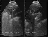
The horse was first seen in the morning and physical examination revealed no abnormalities. Based on the history alone, a thoracic ultrasound was completed (Figure 1).

Figure 1: Ultrasonographic images captured from the thorax of a horse. There is evidence of mild low volume pleural effusion.
As was expected, the sample was purulent with a >250,000 white blood cells per microliter. The cells were fairly non-reactive. The culture was negative for aerobic and anaerobic bacteria. A serum chemistry profile, fibrinogen concentration and complete blood count were performed simply for screening purposes. The results were predictable and included a slight neutrophilic leukocytosis (12,600 total cell/ul with 8200 neutrophils/ul) with no band neutrophils evident. The horse had a normal fibrinogen (150 mg/dl). All other parameters were within normal limits with no concerning tendencies.

Figure 2: Thoracic fluid from a horse with fever of unknown origin.
Treatment and outcome
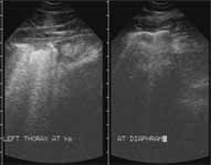
The horse was kept for treatment and observation. She was administered Naxcel™ (ceftiofur sodium) at a rate of 1 mg/pound (2.2 mg/kg), q12h, intravenously via percutaneous injection for 10 days. Temperature was evaluated in the morning and afternoon. As often occurs with pyrexia in horses, the fever was evident in the afternoon, but not the morning evaluation. The horse would vary in the afternoon between 102.1° F (38.9° C) and 103 (39.4° C) with a tendency to decline, but not to resolve, over the course of the 10 day drug regimen. The pyrexia occurred intermittently over the next 8 days and resolved on day 8 after antimicrobial treatment was ceased (18 days from the start of antimicrobial therapy). A recheck ultrasonographic evaluation was performed (Figure 3) on day 28 and indicated extensive resolution of the purulent fluid. The horse went on to do well with no other episodes of pyrexia.

Figure 3: A recheck ultrasonographic image collected from the same horse as Figure 1. Note the near resolution of the pleural pathology. Some small amount of fluid still persists.
Discussion
It has been the author's experience, that a thoracic ultrasound is warranted whenever a horse presents with persistent continuous or intermittent pyrexia and few if any other clinical signs. This can be performed with a 5 megaHz linear ultrasound probe. Clearly thoracic ultrasonographic evaluation is more warranted if there is a purulent nasal discharge or cough, or other more "text book described" clinical signs of pleuropneumonia. Many of the mild or occult cases have small amounts of purulent pleural fluid that has increased echodensity and opacities in the fluid. However, the volume can be small and have a very elevated white blood cell count (Figure 4). A personal review of 48 cases where imaging has been captured indicates that shipping, parturition, or exercise, were evident in many horses before pyrexia developed. In many cases, no specific historical incident was identified.
As is common, in the author's experience with these cases, antimicrobial therapy did not seem to resolve the pyrexia in the short term. There is a tendency to declining temperature and less depression, but less often resolution during the antimicrobial regimen. There seem to be no advantage of applying antibiotics until the pyrexia is completely resolved. That does not seem to be fruitful, reduce the duration of the disease, nor necessary in these cases.
References
Bertone JJ. Management of pleuropneumonia. Proceedings of the Scientific Meeting of the American College of Veterinary Internal Medicine. Chicago. 1999.
Byars TD, Becht JL, Dainis CM. Cranial mediastinal loculation and abscessation as a sequelae to pleuropneumonia in the horse. abs. Proc 5th Amer Coll Vet Int Med Forum, 1987;928.
Rantanen NW, Gage L, Paradis MR. Ultrasound as a diagnostic aid in pleural effusion in horses. Vet Rad 1981;22:211-216.
Rantanen NW. Diseases of the thorax, In: Diagnostic Ultrasound. Vet Clin N Amer 1986;2:49-66.