Diseases of the equine cornea (Proceedings)
Information on various diseases of the cornea in horses.
•Thickness
•< 1mm thick
•Thinnest at center
•0.56 mm
•Exposed Surface
•Epithelium 10-15 cell layers thick
•Stroma
•Bulk of Cornea
•Collagenous fibers (Type 1 and 2)
•Descemet's membrane
•Homogenous membrane of fine collagenous fibers
•"basement membrane"
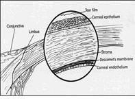
•Endothelium
•Deepest layer
•Pump's fluid out of cornea
•Deturgescent state
•Nerve Supply
•Ophthalmic branch of Trigeminal (V)
•Naked nerve endings : Stimulation = Extreme Pain
•Located just below the epithelium
•Superficial abrasions : Very Painful
•DEEP STROMA
•Nerves are more sensitive to pressure than to painful stimuli
•Metabolism
•Avascular: How does it get oxygen and metabolites
•Peripherally positioned limbic capillaries
•Aqueous Humor
•Tear Film : Major source for dissolved oxygen
Response to Injury
•Epithelial cell mitosis occurs only in basal layer
•Stem cells at limbus
•After injury cells at wound edge enlarge & loose attachments to BM, slide over defect
•0.6 mm/day
•5-7 days for superficial ulcer to heal
•Six weeks are needed to attach the epithelial cell basement membrane to the stroma
•Initially monolayer of cells, but eventually multilayered sheet will cover wound
•Mitosis resumes after wound closure
•Deep Ulceration
•Stromal Cells
•Do not regenerate
•Replaced by Scar tissue
•Fibroblasts
•Accompany blood vessels
•Visible 3-5 days after injury
•Progress 1mm/day
•Deposit collagen fibrils in regenerating stroma
•Disorganized, therefore transparency is decreased
•Endothelium
•Minimal to no mitosis, therefore limited capacity for regeneration
•When cells are lost or damaged, remaining cells enlarge & migrate
DIAGNOSTICS
•Look at orbit, eyelids, pupil and globe position.
•Entropion
•Scraping for Cytology and Cultures
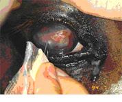
•The cultures should be done before any drops, (drugs contain bacteriostatic agents)
•Topical anesthetics and the handle end of a scalpel blade to scrape.
•Sterile dacron swabs for culture.
•Culture cornea BEFORE applying anything to the eye if overt infection is suspected
•Aerobic
•Fungal
•Fluorescein stain every eye that is PAINFUL!
•Blepharospasm
•Epiphora
•Redness
•Corneal opacities
•Rose Bengal Stain
•Viral Keratitis
•Early Fungal Keratitis
•Assess tear film
•Deep corneal scrapings, at the edge and base of the ulcer to detect bacteria and fungal hyphae
•Obtained with topical anesthesia and the blunt, handle end of a sterile scalpel blade.
•Superficial swabbing cannot be expected to yield fungi in a high percentage of cases.
Corneal Ulceration
•Characterize
•Depth
•Superficial
•Deep (Stromal)
•Descemetocele
•Perforating
•Cause
•Infectious (fungal, bacterial, viral)
•Traumatic
•Immune-mediated
•Duration
•Response to previous treatments
Superficial Ulceration
•Epithelium only
•Foals (Increased incidence compared to adults)
•Corneal sensitivity in foals is very poor.
•Lower tear production in foals compared to adult's
•Painful
Stromal Ulceration
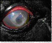
•Epithelium and collagen is lost
•Variation of the clinical picture
•Descemetoceles
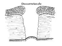
•Epithelium and stroma are not present
•Buldge of DM into the defect
•EMERGENCY!
Corneal Rupture
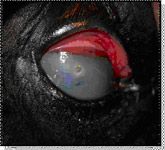
•Iris Prolapse
•Is this an EMERGENCY?
•Owner's Expectations?
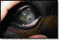
•Cosmetic = Time and $$
•Enucleation?
•Treat as if globe is infected
•Systemic Antibiotics
•Cannot chance bacterial encephalitis
Bacterial Ulcerations
•Gram Positive
•Streptococcus -hemolyticus
•Staphylococcus spp.
•Corynebacterium spp.
•Gram Negative
•Pseudomonas aeruginosa
•Aspergillus
•E. coli
•Treatment Options
•Gram Positive
•Triple Antibiotic
•Solution or Ointment
•Chloramphenicol
•Ointment
•Solution (1 gram vial with 10-15cc of sterile water)
•Cefazolin
•Solution (1 gram vial diluted with 5ml sterile water)
•Ciprofloxacin
•For Staph.
•Treatment Options
•Gram Negative
•Gentocin
•Solution or Ointment
•Tobramycin
•Ciprofloxacin
•Chloramphenicol
•UVEITIS
•Noted with all bacterial ulcerations!
•Melting (Keratomalacia)
•Can occur even if you are on the correct antibiotic(s)
•WHY?
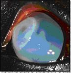
•Complex Healing Process
•Proteinases
•Removal of devitalized cells and debris
•Two major types
•Matrix Metalloproteinases (MMP 2 and MMP9)
•Serine proteinases
•Neutrophil elastase
•Kept in balance by natural anti-proteinases
•a-macroglobulins
•Tissue inhibitors of MMP (TIMPs)
•a-1-proteinase inhibitor
•Bacteria (Pseudomonas and Streptococcus)
•Bacterial Proteinases
•Activate latent corneal MMP
•Proteinase activity in Tear Film
•Increased with ANY ulceration
Anti-Proteolytic Medications
•N-Acetyl Cysteine 5-10%
•MOA: MMP Inhibitor : Chelating Ca
•5 mL 20% Mucomyst in 15 mL artificial tears
•EDTA 0.2%
•MOA: MMP Inhibitor : Chelating Ca
•5ml of sterile water to lavender tube (EDTA)
•If larger tube fill with sterile water until vacuum is gone
•Galardin (Ilomostat®)
•Commercially available
•Antproteinase solution
•Inhibits MMP1, MMP3 and MMP9
•Demonstrated in rabbit model to work very well
•Serum
•MOA: MMP and serine protease inhibitors
•Good for 7-8 days refrigerated
•Cheap!!
•Tetanus Anti-toxin
•MOA: MMP and serine protease inhibitors
•I tend to use 2 anti-proteolytic medications
Fungal Infections
•Aspergillosis and Fusarium
•Common Fungal isolated in horses
•Think Fungal Keratitis if:
•Ulcer is refractory to treatment with topical antibiotics
•Stromal Melting
•Lives or has traveled in warm/humid environment
•Lack of vascularization despite chronicity of ulcer
•Severe blepharospasm despite small size of ulcer
•Eye has been treated with steroids
•Uveitis increases after treatment with antifungals
•Can be VERY frustrating
•Can present with a wide variety of appearances
•Can do whatever they want to – despite appropriate therapy
•Most likely will need surgical intervention
•Lots of PMN
•To small to ingest the hyphae
•Release LOTS OF PROTEASES
•Antiangiogentic factors
•Slow healing
•4-6 weeks minimum
•Topical Treatment
•Natamycin 3.3 % Solution
•Take new bottle (5%) and add 7.5cc Sterile water to make a 3.3% solution
•Produced by Streptomyces spp.
•Normally found in equine conjunctiva/cornea
•Most Physiologically appropriate? (per Dr. Brooks)
•Fluconazole 1%
•Itraconazole 1% with 30% DMSO Ointment
•Nice levels in the cornea
•Miconazole Vaginal Cream
•SSD
•Betadine Solution
•Vericonazole
•Be aggressive Q1-2 hrs if needed
•Treat Uveitis
•Can be severe
•Flunixin meglumine
•mg/kg PO or IV BID
•Atropine Q 4-6 hrs
•Treat with Antiprotease
•Treat with broad spectrum antibiotic
•Neopolybac
•Repeat Cytology and C/S
•If the eye first responds to treatment then deteriorates.
•May have secondary invasion of bacteria
•GOALS OF THERAPY
•Resolve Infection
•HALT Keratomalacia
•Control Uveitis
•Dilate Pupil
•NSAIDS
•Provide Structural Support
•Simple Ulcer
•Neopolybac Ointment
•Uveitis
•Banamine
•Atropine Q8hrs to affect
•Then SID or EOD
•Antiproteolytics +/-
•Re-Check 2 days
•Not Responding
•Cytology, C&S
•Add Chloramphenicol?
•Debridement?
Debridement
•Remove loose "lips" of epithelium that are non-adherent
•Topical anesthetic
•Dry Q-tip
•Rub cornea (utilize friction) in a circular, curvilinear fashion out from edges of wound
•Replace Q-tips often to keep dry
•Superficial ulcers only
•CHEMICAL CAUTERIZATION
•3% to 7% Iodine
•Good for Fungal Keratitis
Melting Ulcer
•Cytology, C&S
•Debridement
•Remove Keratomalacia
•Chemically Cauterize
•SPL
•Atropine Q 4 hrs to affect
•Chloramphenicol solution
•Triple antibiotic solution
•Diflucan QID
•Tetanus Antitoxin
•EDTA (If you wish)
•NSAID
•Banamine Q 12 hrs
•Equioxx?
•Cytology, Culture and Debride
•Chemically Cauterize
•SPL
•Chloramphenicol Q 2 hrs
•Triple Antibiotic Q 2 hrs
•Atropine Q 6 hours to affect
•Diflucan Q 4 hrs
•Serum Q 2 hrs
•NSAID
•Banamine
•Cytology, Culture and Debride
•Chemically Cauterize
•SPL
•Chloramphenicol Q 2 hrs
•Triple Antibiotic Q 2 hrs
•Atropine Q 6 hours to affect
•Diflucan Q 4 hrs
•Serum Q 2 hrs
•NSAID
•Banamine
Stromal Ulceration
•Cytology, C&S
•Place SPL
•Chloramphenicol Q2
•Neopolybac Q2
•Atropine Q 6 Hrs
•Banamine
•Serum Q 2 hrs
•EDTA Q 2 hrs
•Diflucan QID
•Stroma rigidity should show rigidity after 48 hrs of treatment
•If not = Flap
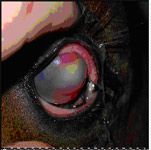
Stromal Abscess
•White to yellow opacities within the cornea
•Accumulation of inflammatory cells & products, +/- infectious agents
•Variable stromal involvement (superficial, deep, full-thickness)
•Usually overlying epithelium is intact, but not always
•Surgical
•Minimal Vascularization
•Progresses
•Cannot control uveitis
•Worried about vision loss
•Penetrating Keratoplasty
•Medical
•Chloramphenicol
•Itraconazole and DMSO
•Ciloxan
•NSAID
•Atropine
•BUY TIME FOR THE CALVARY (Vessels)
•Penetrating Keratoplasty
•Full thickness resection
•Remove abscess
•Place and suture full thickness corneal graft
•Cover with conjunctival graft
Fungal Keratitis
•SPL
•Serum 6x daily
•Ciloxan 6x daily
•Natamycin 6x daily
•Miconazole 6x daily
•Atropine 1x daily
•NSAIDS
•Banamine
•Expect 3 months worth of treatment
•Squamous Cell Carcinoma
•Most Common Tumor of the Eye
•Lesions are pink and begin at the limbus
•Extending to the cornea they can appear like cauliflower
•Painful at times
Squamous Cell Carcinoma
•Treatment consists of lamellar keratectomy with adjunctive tx
•Cryotherapy
•Beta-irradiation (Sr 90 )
•Radiofrequency hyperthermia
•CO 2 laser ablation
•Treat wound bed as an ulcer, cover if possible (conj flap)
•Squamous Cell Carcinoma
•Medical Therapy
•5-fluorouracil
•Mitomycin-C
•These are usually just palliative
•Mitomycin
•Order from Wedgewood Pharmacy
•Give 3-4 x daily for 3-4 weeks
•Cost = 100-150$ a week
Eosinophilic Keratitis
•Inflammatory cells with eosinophils on cytology
•Unknown etiology (allergic or parasitic??)
•Treat is often prolonged, taper should be slow
•Topical antibiotics
•Topical corticosteroids
•If no stain uptake
•Topical mast cell stabilizers (I do not like)
•Cromolyn
•Systemic NSAIDs\
•Systemic Steroids if stain uptake
•10mg PO SID for 5-10 days
•+/- Cyclosporine
•In KY see in Late Summer