Managing infectious equine respiratory diarrheal disease (Proceedings)
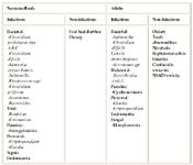
Salmonella enteriticus, Neorickettsia risticii (Potomac Horse Fever), Clostridium difficile and Clostridium perfringens are most commonly associated with infectious diarrhea in adults. Foals can have a variety of agents including viral causes and bacterial such as Lawsonia intracellularis.
Rule Outs for Infectious Diarrheal Diseases
Salmonella enteriticus, Neorickettsia risticii (Potomac Horse Fever), Clostridium difficile and Clostridium perfringens are most commonly associated with infectious diarrhea in adults. Foals can have a variety of agents including viral causes and bacterial such as Lawsonia intracellularis. However, it is estimated that less that half of the cases of enterocolitis, an etiologic agent is ever identified in acute diarrhea of the horse. Other causes include parasisitism with larval cyathostomiasis a suspected culprit in adults and several verninous parasites in foals. Non-infectious causes are likley under appreciated and under recognized and these primarily include dietary changes (composition or quantity), toxicity (e.g., heavy metals, phenylbutazone or cantharadin toxicosis), sand and antibiotic-associated diarrhea. In all the of the non-infectious causes, horses can become febrile and toxic due to the severe secondary effects associated with massive changes in flora and secondary endotoxemia due to changes in composition of gram negative flora. Nonetheless, any febrile, diarrheic horse should be treated as infectious and biosecurity precautions instituted.

Salmonellosis
Like humans, initial infection is likely foodborne in the adult, but the role of maternal-to-foal transmission during neonatal life has been largely uninvestigated. Furthermore, the role that foal carriage plays in environmental maintenance and cycling is largely unappreciated. Salmonellosis is considered by some to be the most common cause of infectious enterocolitis in adult horses, although a recent report from California suggested that Clostridium difficile might be more common in that state. The reported prevalence of infection with Salmonella has been variable, ranging from less than 1% (NAHMS) to 70% with higher prevalence of Salmonella associated with 1) the presence of clinically affected horses; 2) method of detection); and 3) climates with warmer months of the year (summer and autumn) associated with outbreaks in Florida and Fall and winter in North Dakato. According to Cohen (1997, 1998) the risk factors for equine enteric salmonellosis include 1) transportation, 2) change in diet, 3) antimicrobial treatment, 4) surgery, 5) common nasogastric tubes, 6) wet and dark conditions, and 7) other gastrointestinal disorders (e.g., impaction colic). Foals are at risk for bacteremia/sepsis caused by Salmonella. Mares are commonly considered to be the source of infection, although are usually without clinical signs of disease (Madigan). It has been proposed that the underlying mechanisms for known risk factors is a change in the intestinal microflora. In the United States, Salmonella enterica serovar Newport is the serovar most implipaced in large hospital outbreaks. This serovar is considered by CDC to be under epdemic spread in animals and humans.

Comments on Laboratory Findings/Ancillary Testing
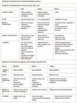
Diarrhea is the most commonly observed clinical sign. Not all horses with salmonellosis enterocolitis will develop diarrhea nor will they be febrile. Presence of fecal leukocytes may help to attribute any colitis to an invasive pathogen. For fecal culture, collect several grams of feces. Rectal swabs are insensitive. Transport using suitable transport media (e.g., Ames aerobic culture media) and keep cold. Double wrap all samples and wipe off outside of sample with 10% bleach before sending. Samples can be transported in selenite broth if processed within 24 hours of collection. Five samples for culture and three fecal samples for PCR must be submitted before a horse or foal can be declared negative. For fecal PCR, this technique appears to be highly sensitive and a positive result likely carries greater importance in a foal than adult horse with diarrhea. All foals should have a blood culture performed. This is particularly useful in foals less than 1 month of age, as young foals with intestinal Salmonellosis are frequently bacteremic.

Comments on Treatment
Antimicrobials are controversial in adult Salmonellosis but are frequently recommended for all juvenile horses with enterocolitis. Antimicrobials that have been used frequently to treat salmonella infections in foals include chloramphenicol, ceftiofur, and enrofloxacin. Recent evidence does demonstrate the transfer of antimicoribial resistance genes between E. coli and Salmonella in horse strains, however, cattle and poultry are still the major carriers of multiresistantce genes in Salmonella. Nonetheless equine salmonella isolates can carry their own unique resistance genes and a specific mutation has been identified that leads to fluoroquinolone resistance. The S. enterica serovar Newport this is undergoing expansion across the U.S. has been characterized on the basis of its resistance. Thus, based on recent work with animal and human strains, antimicrobieals should be avoided in all cases of animal salmaonellosis except where evidence of neonatal infection in foals indicates systemic spread (osteomyelitis, lung, liver) and the treatment is therefore life saving.

Clostridial Infections
Epidemiology. According to data from California and Colorado the risk factors for clostridial enterocolitis include 1) age, 2) use of antimicrobials, 3) management factors, and 4) hospitalization. C. difficile infection is more likely associated with acute colitisi in mature horses which have been treated by antibiotics. Recent studies demonstrate that foals, although they are susceptible to infection and idsease, are more resistant. Pathogenesis. Clostridia difficile has five toxins, although the disease causation is thought to rely on the production of toxins A and B . The evidence to date is unclear of the exact contribution of either toxin A or B mediating pathogenesis of C. difficile diarrhea. Research suggests that the formation of the classic pathologic lesion, the pseudomembrane is a result of the combined actions of toxin A, toxin B, interleukin-8. Classically, toxin A is considered an enterotoxin and lethal while toxin B is a virulent cytotoxin. It appears that most horse isolates have both toxins.
Diagnostic Testing. For clostridial enteropathogens, 20 to 30 ml of feces submitted in an anaerobic transport device, is needed to demonstrate the presence of toxin. Some laboratories offer immunoassays to detect toxin(s) produced by C, difficile and the enterotoxin of C. perfngens.Feces should be examined by sedimentation for sand and microscopically for increased fecal leukocytes. A Gram stain may be useful in detecting Clostridial diarrhea (long Gram positive rods). Detection of cyathostomes larvae by direct examination of feces. If cantharadin toxicosis is suspected the feces should be examined for parts of Epicauta beetles. The hay should also be checked for beetles.
C. perfringens Diagnostic Update Recovery of Cl. perfringens from diarrheic foals is also of questionable significance as the organism is commonly present in the feces of healthy foals, particularly Cl. perfringens biotype A. The genes that encode the Clostridial toxins can be amplified and categorized using PCR techniques. All Cl. perfringens isolates contain an alpha toxin. Further separation into biotypes A through E is based on identification of additional toxins produced by the bacteria. The exception is a Cl. perfringens isolate that is yet to be biotyped, but contains the alpha toxin and a 2 toxin. The latter toxin has similar biological activity to the toxin, but has no significant amino acid homology with that toxin. This organism has been isolated from both adult horses and foals with diarrhea. Samples should be shipped directly to a diagnostic laboratory immediately or transported on ice and shipped overnight. The traditional method for toxin identification within intestinal contents or fecal samples is through mouse inoculation. Use of PCR has largely supplanted biological assays. However, one must remember that identificaiton of a toxin by this manner is correlative. Any large outbreak should also be biotyped. The PCR techniques involves culture of C. perfringens from the sample and then toxin testing.
Therapy: Metronidazole should be used for clostridial colitis (15-20 mg/kg; q. 8 to 12 hrs; PO), although approximately one third of recent Clostdium diffrcile isolates from California were thought to be resistant. Resistance is relative however. Infection in humans demonstrates little difference in relapse rate based on antibiotic therapy.
Lawsonia Infection
Lawsonia intracellularis infection causes a specific syndrome in foals characaterized by protein loss, internittent diarrhea, weight loss, and ill thrift. Foals are usually older between 3 and 8 months. Outbreaks can occur on a single farm and can recurr at those locales yearly. Erythromycin has been advocated as the treatment of choice, but recent publications indicate tetracyclines or chloramphenical as alternatives. Diagnosis can be difficualt and relies of eihter a positive ELISA testing for antibody to Lawsonia intracellularis and/or PCR performed on feces. Since this disease is sporadic, equine isoltes have been difficult to fully characterize and data regarding diagnostic techniques is difficualt to quantify.
Equine Monocytic Ehrlichiosis or Potomac Horse Fever
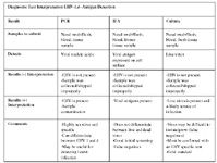
Diagnosis. Both indirect fluorescent antibody (IFA) and enzyme linked immunosorbent assay (ELISA) test formats have been developed to detect antibody for N. risticii, however, most diagnostic laboratories rely upon the IFA. Paired serum titers must be evaluated; single titers are useless for confirmatory testing for EME. A value of 1:80 has been proposed as positive, however, utilizing PCR techniques, a few naturally infected horses were actually positive in the face of lower titers. Experimentally infected horses experience a large rise in titer between 4 and 10 days into the onset of clinical signs; a 4-fold rising titer can be demonstrated often within the first week. The IFA does not differentiate titers that are the result of vaccination or previous infection. A high number of false positive results also afflict the IFA format. Polymerase chain reaction performed on buffy coat or feces is a sensitive way to detect N. risticii antigens. In experimentally infected animals, PCR performed on buffy coat or feces was more sensitive than culture. However PCR has proven somewhat less sensitive than culture for clinical specimens. This slightly decreased sensitivity may be due to specimen handling. Under appropriate specimen handling, PCR offers a sensitive detection technique with minimal delay. Furthermore, in clinical specimens, detection by PCR on blood is more sensitive than detection in feces. This latter finding may also be due to specimen handling and quality, because large numbers of N. risticii are shed into bowel lumen in epithelial cells. In untreated horses, N. risticii can be detected for up to 30 days post-infection by PCR. Comparison of IFA to PCR detection, horses may be PCR positive even when titers are still low, <1:20. Thus testing early during the stage of disease, before treatment, will likely yield positive clinical specimens.
Therapy. Tetracyclines still remain the drugs of choice against N. risticii infection. Tetracycline should be administered during clinical illness in ehrlichial infections. Similar to other Ehrlichia such as Anaplasma in cattle and Ehrlichia in dogs, treatment of horses during the incubation period will delay clinical signs, but illness, albeit somewhat less severe, still may occur. Treatment with oxytetracycline (6.6 mg/kg, IV, q 12 hrs for 5 days) in horses displaying clinical illness due to N. risticii results in rapid response to treatment within 24-48 hours.