- dvm360 August 2020
- Volume 51
- Issue 8
The ABCs of veterinary dentistry: W is for waiting to treat
When it comes to dental care, sometimes the best course of action is no action at all.
I can vividly remember back in 1974 when Dr. Wiggins, one of my professors at Auburn University College of Veterinary Medicine, advised us students to wait until a deciduous “double” tooth falls out by itself. Fast forward many years, and I now know that postponing the extraction of persistent primary teeth often causes harm to my patients.
Waiting to perform optimum dental care is sometimes the treatment of choice and at other times is a bad idea. This article covers some of those situations where waiting is the best option. A second article will outline conditions for which waiting is just wishful thinking.
Plaque and tartar without inflammation
Periodontal disease arises from gingivitis caused by plaque irritating the gingiva. When left to accumulate, plaque calcifies into rough calculus (tartar), which allows more plaque to collect on the crown. In predisposed dogs and cats, plaque inflames the gingiva (gingivitis) and eventually progresses to loss of support (periodontal disease).
Periodontal disease cannot occur without gingivitis, but gingivitis can occur without progressing to periodontal disease. Regardless, once calculus contacts the gingiva and inflammation is noted, it is time for an anesthetized professional oral hygiene visit that includes dental scaling, irrigation, and polishing; tooth-by-tooth probing and full-mouth intraoral imaging; and treatment for any underlying disease discovered. Waiting will only increase patient discomfort and the extent of disease (Figure 1).
If calculus is clinically present but inflammation is not, the professional oral hygiene visit can wait either until inflammation becomes apparent or a year has passed from the time when the last professional oral hygiene visit, to remove the accumulated plaque and calculus.
Stage 2 external root resorption confined to the subgingiva
External root resorption can occur anywhere on a cat’s or dog’s tooth but starts in the periodontal ligament space. Many root resorptions are discovered via intraoral radiographs obtained during the anesthetized professional oral hygiene visit. Even though external root resorption is considered progressive, immediate extraction is not necessary when the resorption is shown clinically and radiographically to be located far below the gingival margin in the apical third of the tooth. Once external root resorption is exposed to the oral cavity, bacterial contamination of dentin and pulp results in painful inflammation. In those cases, extraction is the treatment of choice (Figure 2).
Internal root resorption is a different story in that by its nature the pulp is infected and considered painful. For internal root resorption, waiting is not an option. Root canal therapy or extraction are the treatments of choice.
Supernumerary and malpositioned teeth
Extra teeth in large-breed dogs often can be accommodated without causing harm or requiring extraction. Small-breed dogs and most cats do not have this luxury. Supernumerary teeth in small- and medium-sized breeds can cause tooth crowding, leading to gingivitis and periodontal disease resulting from the accumulation of food and oral debris between the teeth irritating the gingiva. Immediate extraction of the tooth (teeth) causing crowding is indicated in these cases (Figure 3).
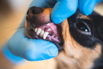
Not all teeth erupt in normal positions. Malpositioned teeth may be secondary to birthing trauma, inherited defects, or trauma after birth. The decision to treat or wait to move or extract abnormally positioned teeth should be made after an examination that includes probing and intraoral radiographs. If the malpositioned tooth is functional and not causing discomfort, waiting to extract is the best option (Figure 4).
Generally, whether to extract abnormally located teeth can present a conundrum. If there is no obvious mucosal penetration causing discomfort, a wait-and-see approach is appropriate.
Abrasion and attrition
Chronic abrasion (i.e., tooth wear caused by contact with a nondental object, such as from self‐grooming or chewing on tennis balls) and attrition (caused by contact of a tooth with another tooth from misaligned opposing teeth) may result in excessive wear and direct or indirect trauma to the pulp. Repeated low‐grade trauma stimulates odontoblasts to produce tertiary (reparative) dentin for repair and protection. Tertiary dentin often appears as a reddish‐brown shiny spot in the center of the worn surface.
As long as the rate of wear is gradual, reparative dentin production will keep up with loss of tooth structure without causing pulpal exposure. When the rate of wear is faster than the rate of tertiary dentin production, the pulp becomes exposed, leading to bacterial invasion pulpitis and eventually pulp necrosis. Probing the worn area with an explorer and radiographic examination will help evaluate endodontic and periodontal involvement of worn teeth to see whether therapy is indicated. In cases of pulp exposure, treatment (root canal therapy or extraction) is indicated because pain and infection usually result (Figure 5).
Articles in this issue
about 5 years ago
Gene editing in animals: Miracle or madness?about 5 years ago
Compassion-First adds 43rd veterinary practiceabout 5 years ago
First diagnostic test for canine IBD now availableabout 5 years ago
FDA approves generic carprofen chewable for dogsabout 5 years ago
When cats get hangry…about 5 years ago
Why we need more veterinary colleges at HBCUsabout 5 years ago
DNA debunks age-old ratio for dog years to human yearsabout 5 years ago
Quiet, please!Newsletter
From exam room tips to practice management insights, get trusted veterinary news delivered straight to your inbox—subscribe to dvm360.


