Treating UTIs with fluoroquinolones: A case study (Sponsored by Pfizer)
Florie is a 4-year-old, spayed female Labrador-shepherd crossbred dog. Until six months ago, Florie's medical history was unremarkable. At that time, she was diagnosed with a bacterial urinary tract infection (UTI) based on the presence of hematuria and pollakiuria; she received 500 mg cephalexin orally once daily by the referring veterinarian for five days.
Florie is a 4-year-old, spayed female Labrador-shepherd crossbred dog. Until six months ago, Florie's medical history was unremarkable. At that time, she was diagnosed with a bacterial urinary tract infection (UTI) based on the presence of hematuria and pollakiuria; she received 500 mg cephalexin orally once daily by the referring veterinarian for five days. When the UTI was observed again by the referring veterinarian three months later, Escherichia coli was cultured but sensitivity results were not obtained. Florie was given 50 mg gentamicin subcutaneously three times daily for 10 days.
Three months later, Florie was referred to the University of Georgia Veterinary Medical Teaching Hospital (UGA-VMTH). At this time, she was exhibiting a recurrence of the same clinical signs (i.e., pollakiuria, dysuria, and hematuria). During this exam, the owner reported that Florie had actually experienced pollakiuria and hematuria intermittently for six months and had recently increased her water intake. Florie weighed 44 lbs and her physical examination findings were normal except that she was reluctant to allow caudal abdominal palpation and her urinary bladder appeared small. Because of her size, body conformation, and reluctance for abdominal palpation, Florie's kidneys could not be palpated. Rectal palpation of the distal urethra was normal.
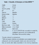
During Florie's exam at UGA-VMTH, a complete blood count (CBC), serum chemistries, urinalysis, urine culture, abdominal radiographs, and ultrasonogram were performed. The results of the CBC and serum chemistries were normal and the urinalysis and urine culture results are listed in Table 1.

Table 1: Results of Urinalysis at UGA-VMTH
The abdominal radiographs revealed normal abdominal viscera. Florie's kidneys were normal-sized but irregularly shaped. The urinary bladder was small. No uroliths were visible. On the ultrasonogram, her kidneys were normal-sized, but medullary echogenicity was irregular and abnormal bilaterally. The renal pelves were mildly dilated bilaterally. A 2-cm polyp was present in the body of the urinary bladder. No uroliths were visible. Based on the physical examination and laboratory test results at UGA-VMTH, the clinical diagnosis was UTI with renal involvement and polypoid cystitis. This clinical course (three successive presentations over six months) is, unfortunately, not uncommon for UTI. This article evaluates these three presentations at zero, three, and six months of Florie's illness.
Initial identification of UTI
Canine UTIs generally involve the lower urinary tract (bladder and urethra), where the inflammation caused by the infection produces obvious clinical signs such as pollakiuria, stranguria, and dysuria. These findings in Florie, typical of lower tract disease in general, led to the initial suspicion of a bacterial UTI. However, other causes of lower urinary tract inflammation, such as cystouroliths or neoplasia, may coexist with a UTI or cause lower tract signs in the absence of bacteria. In this case, the presumptive diagnosis of a bacterial UTI was reasonable because it's a common cause of lower tract signs in dogs, particularly young or middle-aged dogs with no other findings.
Origin of the infection
Most bacterial UTIs are caused by intestinal or cutaneous flora that ascend through the urethra to the bladder. The most common bacterial pathogens associated with UTIs in the dog are E. coli (as later found to be the case in Florie) and Staphylococcus, Streptococcus, Enterococcus, Enterobacter, Proteus, Klebsiella, and Pseudomonas species.
Common contributory causes of UTIs were apparently not factors in Florie, such as urethral catheter use, conditions that would cause local inflammation and prevent efficient urine flow from the bladder (e.g., prostate enlargement in males or uroliths), and urinary tract surgery. Nonetheless, a presumptive diagnosis of a UTI was made. The main reservoir for such a UTI is the bacterial flora of the gastrointestinal tract—this was presumed to be the source in Florie's case.
Analysis of Florie's initial therapy
For initial treatment of a simple UTI without known complications, it's appropriate to treat empirically and presume (for the first episode only) that the UTI is limited to the lower urinary tract. Most bladder infections are a superficial mucosal infiltration at an anatomic site where antibiotics can be easily delivered. Generally, products such as potentiated penicillins or cephalosporins are used as first-line antibiotics and fluoroquinolones reserved for severe, resistant, or recurrent infections where culture and sensitivity results are available.
Because E. coli is the most frequent urinary tract pathogen, the empirical drugs of choice are trimethoprim-sulfa or a fluoroquinolone in the absence of bacterial culture results. Because of safety, the latter is preferred as a first choice by some. Initial therapy for a bacterial UTI in this setting should last 10 to 14 days. For reasons that are unclear, a subtherapeutic dosage of cephalexin (10 mg/lb once daily) was maintained for only five days by the referring veterinarian. While bacterial UTIs in people sometimes resolve with three to five days of low-dose therapy, such a short-term approach is unlikely to eradicate a bacterial UTI in veterinary patients because the infection is usually well established before the animal presents to a veterinarian. A therapeutic dosage of cephalexin might have been effective initially in this case, but it would generally not be the drug of first choice for UTI caused by E. coli.
Furthermore, the signs observed on initial presentation, which localized involvement to the lower urinary tract, did not identify the cause of the inflammation or preclude upper tract involvement or an unidentified complication (or prostatic involvement in a male dog). The diagnosis of lower tract infection was a presumptive diagnosis only. Florie should have been reevaluated one to two weeks after cessation of therapy by urinalysis and, preferably, urine culture, but this was not done upon her initial evaluation.
Visit two: Recurring UTI
The referring veterinarian evaluated the UTI again three months after the initial episode. Recurring UTIs can be classified into two categories: relapses and reinfections. Relapses are infections caused by the same bacterial species that occur within several weeks of the cessation of treatment. In Florie's case, it's likely that her inadequate antibacterial treatment failed to eliminate the organism. Relapses may result from using an improper antibiotic, dosage, or duration of therapy as in Florie's case; the emergence of drug-resistant pathogens; or failure to eliminate factors that alter normal host defense mechanisms, such as polyps. Relapses may also result from deep tissue involvement (e.g., kidney and bladder polyps) or, in male dogs, from chronic prostatic infections.
Recurrent UTIs may also result from reinfection. In such cases, initial antibacterial treatment eradicates the first infection, but another bacterium subsequently infects the urinary tract. Reinfections generally occur when host defense mechanisms remain compromised. Though possible, this was not suspected in Florie.
Therapy of Florie's recurrent UTI
The renewed presence of clinical signs at three months and the initial choice of a subtherapeutic antibiotic dose made relapse the most likely cause of Florie's recurrent UTI. During the three-month exam, E. coli was cultured, but sensitivity results were not obtained. Florie was given 50 mg gentamicin subcutaneously three times daily for 10 days.
Choice of empirical therapy can be based on the identity of the infecting organism. As previously mentioned, this would generally be trimethoprim-sulfa or a fluoroquinolone for E. coli. Pseudomonas infections may be empirically treated with tetracycline. For Proteus, Staphylococcus, and Streptococcus species, amoxicillin is often a first choice. For Enterobacter and Klebsiella species, a fluoroquinolone is often the drug of choice. Thus, the choice of gentamicin was not consistent with the above recommendations. It's likely that the organism was sensitive to gentamicin, but the therapy should have lasted longer. Unfortunately, the chosen antibiotic limited the duration of therapy because aminoglycosides add the potential risk of nephrotoxicity, especially when given for 10 days or longer. Once-daily dosing with aminoglycosides is generally less nephrotoxic than more frequent administrations, particularly when the duration of therapy is 10 to 14 days. However, given the long duration of therapy required to clear renal infections, nephrotoxicity becomes a concern no matter how aminoglycosides are administered. A more reasonable choice would have been a fluoroquinolone, with therapy lasting 14 to 21 days or longer.
Therapy for a recurrent UTI involves proper antibiotic selection and a urine culture after three to five days of therapy to verify sterility. The absence of bacteria on a urine sediment exam is not a reliable measure for detecting urine sterility. Interestingly, due to the gradual nature of the healing process, the urine sediment may be abnormal at this time, showing both pyuria and hematuria. For these reasons, a urine culture alone is indicated for this recheck after three to five days of therapy. Florie should have been reevaluated through urinalysis and urine culture one to two weeks after therapy cessation to verify treatment success. Unfortunately, this was not done at this time.
Visit three: Another recurrence of UTI
Florie presented to the UGA-VMTH three months later, approximately six months after initial presentation to the referring veterinarian. Florie exhibited the same clinical signs—pollakiuria, dysuria, and hematuria. This time, however, there was a complaint of polydipsia; in addition, polypoid cystitis was confirmed and renal structural abnormalities were noted on radiographs and ultrasonography.
Complicated vs. uncomplicated UTI
A UTI is considered complicated when an underlying structural or functional abnormality in the host's defense mechanisms is present, making it more difficult to treat than a simple UTI, which usually resolves soon after appropriate antibiotic treatment is initiated. It is usually difficult to eliminate the clinical signs of complicated UTIs with a single course of antibiotics because signs either persist or recur shortly after antibiotic withdrawal. Because male dogs generally experience fewer UTIs than female dogs, any UTI in a male dog should be considered complicated because prostate tissue involvement is likely.
Florie now has evidence of complicating factors that make proper management of a UTI problematic: the infection has been recurrent and a bladder polyp is present. While its location in the body of the bladder and Florie's young age make an inflammatory polyp likely, it complicates therapy. An infected polyp is a deep-seated tissue infection that requires longer-term therapy and an antibiotic with good tissue penetration. Also, the dog has evidence of renal involvement: low urine specific gravity, irregular renal margins, dilated renal pelves, and abnormal renal medullary echogenicity. These changes likely mean that the urinary antibiotic concentration is suboptimal because the urine is dilute.
Localization of the infection
While bacteria may invade the kidney via the bloodstream, most renal infections occur when bacteria ascend to the renal pelvis from the ureter and subsequently invade the medulla. Most upper urinary tract infections are bilateral because colonization factors of certain bacteria, especially E. coli, aid their ascension.
With recurring or complicated UTIs, the question to address is whether the UTI involves the kidneys. Clinicians can do this by relying on clues that help localize the infection to the upper urinary tract. While researchers in human medicine are investigating the value of measuring tubular cell enzyme activity in the urine, serum C-reactive protein, and serum antibody response to bacteria, the results are inconclusive and these tests can't be recommended.
Localization of an infection to the kidney requires an astute clinician. Infections involving the upper tract often produce systemic signs of fever, leukocytosis, fatigue, inappetence, and lumbar pain, especially on renal palpation. A urinalysis may reveal casts, especially leukocyte casts. Survey radiographs may reveal small kidneys that are irregularly outlined with chronic renal infection; with acute kidney infections, the kidneys are usually normal. An intravenous pyelogram or renal ultrasonography may demonstrate the dilated collection system that characterizes renal infection. Decreased urinary concentrating ability—a measure of medullary function—may help identify renal involvement because bacteria tend to localize to the renal medulla, frequently interfering with the concentrating process.
In Florie, dilute urine, dilated renal pelves, irregular renal margins, and changes in renal medullary echogenicity upon her evaluation at UGA-VMTH suggest that the bacterial UTI had ascended to the kidneys.
Intermittent episodes of acute renal infection with clear signs may occur in some patients with chronic renal infection. But long-standing infections sometimes have a less dramatic clinical presentation, making them difficult to diagnose. Unfortunately, for poorly understood reasons, chronic renal infections may be silent, especially in elderly cats and in animals given high levels of glucocorticoids. Many renal infections in people are similarly covert with no clinical signs at all. Routine urinalysis and urine culture in elderly animals and those given high levels of glucocorticoids will help to identify these infections.
Clinical impact of upper UTIs
Obviously, by gaining access to the entire urinary tract and bloodstream within the kidney, these bacterial pathogens have the potential to seed other tissues. Tissues that may become secondarily involved include all genitourinary structures, cardiac valves, lungs, bone, and nervous tissue, but literally any tissue can be affected.
For the clinician, limiting renal damage is a primary concern. Toxic byproducts of bacterial metabolism may damage the renal medulla, interfere with the urinary concentrating mechanism and tubular handling of electrolytes, and obstruct tubular flow, leading to the retention of nitrogenous wastes. Immunologic reactions also play a role in the progression of renal injury from bacterial infection. While nonspecific inflammation and specific immunity directed against bacterial pathogens are theoretically protective, renal tissue is often an innocent bystander. Inflammatory mediators released by leukocytes are toxic to tubular and interstitial cells, contributing to tubulointerstitial fibrosis, which is often seen with advanced renal infection. Furthermore, bacterial infection may expose renal antigens and stimulate an immunologic reaction against self-antigens within the renal parenchyma. Once set in motion, this latter mechanism can continue to destroy renal tissue for prolonged periods after the kidney is sterilized. For these reasons, it is important to rapidly deploy effective therapy to limit renal damage; recurrence and relapse are possible and highly undesirable. Regardless of the specific mechanisms involved—whether bacterial toxins, inflammatory byproducts, or immune mechanisms directed against self-antigens—renal infection is now recognized as a cause of progressive renal disease.
UTI with renal involvement
In Florie, the recurrent clinical signs, evidence of renal involvement, presence of a bladder polyp, and initial choice of subtherapeutic antibiotic doses again make relapse the most likely cause of the recurrent UTI. Furthermore, due to the presence of a renal infection and polypoid cystitis, an antibiotic with good tissue penetration was essential.
Most bladder infections involve superficial mucosal infiltration at an anatomic site where antibiotics can be easily delivered. Veterinarians are accustomed to relying on high urine concentrations of antibiotics to enhance the efficacy of therapy directed against bacteria in the lower urinary tract. This principle doesn't apply to renal infections because bacteria reside in the medulla, outside of the tubular lumina. Consequently, bacteria will be exposed to tissue, not urinary, levels of antibiotics. The same is true of prostatic infections in males. Renal infection is a deep parenchymal infection where antimicrobial delivery is limited and natural host defenses are ineffective due to the hostile environment. Renal infection and polypoid cystitis require a more intensive, prolonged course of therapy. Renal infections are characterized by intense inflammatory responses and bladder infections by limited inflammation.
Successful management of Florie's UTI
On Florie's visit to UGA-VMTH, the organism cultured from Florie's urine was sensitive only to ampicillin, cephalexin, gentamicin, and enrofloxacin using Kirby-Bauer disk sensitivity testing. Disk sensitivity testing should accurately predict drug efficacy with deep tissue infections, such as Florie's polypoid cystitis and renal infection. Although other fluoroquinolones were not tested, it was assumed that this organism was also sensitive to marbofloxacin, which is often the case with fluoroquinolones, and Florie received 75 mg marbofloxacin once daily for six weeks. This fluoroquinolone is excreted in high concentrations in the urine of dogs, and good tissue penetration in the kidney and bladder polyp was anticipated. Sterility of the urine was confirmed by urine culture five days later and again at three weeks of therapy. Fourteen days after cessation of therapy, Florie's urine was sterile and the bladder polyp was smaller. Although a remnant of the polyp remained, it was decided to follow up with urine cultures at three-to six-month intervals.
A year later, Florie's urine remains sterile, her renal function is stable, and no further changes have been noted on renal ultrasound. However, it will be important to perform rechecks every six months to follow the progress of her kidney disease and make sure her UTI doesn't recur. Complicated UTIs usually recur when animals, such as Florie, fail to respond to short-term empirical therapy. These cases can have a happy ending, but this requires a thorough evaluation of the patient, routine use of urine culture and sensitivity testing, and long-term therapy with a properly chosen antibiotic.