Triaging colic patients (Proceedings)
Triaging colic patients.
Signalment and history
• The history can be very brief in order to speed up the examination process
• The history can be conducted after the initial exam for horses with active signs of colic.
• The most critical pieces of the history:
o Treatments already administered
o Any known reactions to medications
o Duration of colic
o Severity of colic
Examination of the horse with colic
• Physical examination (TPR, peripheral pulse quality, mucous membrane color, capillary refill time, auscultation of the chest and abdomen)
• Assessment of dehydration
• Listen for the frequency and quality of gut sounds (Over 1-minute, gut sounds should be present in the upper and lower quadrants of both sides of the abdomen). Specific gut sounds include:
o Opening of the ileocecal orifice (sounds like emptying of a drain)
o Sand in the ventral colon (sounds like sand within a paper bag as you slowly turn it over)
o Short, sharp tinkling sounds such as those you experience with 'GI upset.'
• Rectal findings. Normal findings include:
o Bladder
o Reproductive tract
o Ventral band of the cecum on the right
o Aorta dorsally
o Left kidney
o Nephrosplenic ligament
o Spleen
o Pelvic flexure (or doughy colon on the lower left quadrant)
• Nasogastric reflux (up to 2L is normal)
• Abdominocentesis (normal: TNCC < 10,000 cells/μl; TP < 2.5g/dl)
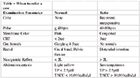
When to refer to a case(see table)
• Refractory or unrelenting pain
• Lack of response to therapy
o Be thinking of referral the second time you go out to see a patient
o Evidence of endotoxemia (consistently elevated heart rate, congested gums, prolonged capillary refill time)
o A finding inconsistent with a simple colic, such as excessive reflux (> 2-5L), a distended viscous, tight band, or extensive impaction on rectal examination, a serosanguinous abdominocentesis
Causes of nasogastric reflux:
• Pyloric obstruction
• Small intestinal obstruction or strangulation
• Nephrosplenic entrapment of the large colon
• Occasionally with large colon volvulus
• Anterior enteritis
Causes of tight bands:
• Large colon displacement or volvulus
• Grossly distended cecum
• Mesentery under tension
• Uterine torsion
Causes of abnormal abdominal taps:
• Small intestinal compromise (strangulation or prolonged simple obstruction)
• Enteritis
• Large intestinal compromise (prolonged simple obstruction)
• Splenic tap

How to prepare a patient for referral
• Encourage your clients to pre-plan for an emergency:
o Which horses will they consider referring based on emotional and financial factors?
o Who will make decisions if the owner is away?
o Consider insurance on those horses for which colic surgery would be a consideration
o Is there a truck a trailer available consistently?
• If the horse is severely dehydrated, consider placing a catheter and bolusing fluids (at least 20L)
o If reflux was obtained from a nasogastric tube, tape the tube in place. This can prevent a ruptured stomach. Place a rubber glove with a finger cut off over the end of the tube to act as a valve.
o Provide enough analgesics/sedatives for the duration of the trip.
o The horse should be confined in the trailer to prevent attempts at recumbency and rolling en route.
o Call the referral hospital prior to the horse's departure
o Have the referral veterinarian talk to the owner about the costs
o Send a copy of the examination findings and drugs administered