Veterinarians must be aggressive to detect distal limb lameness in sport horses
Rigorous evaluation techniques and diagnostics are key for veterinarians to identify lameness early and for successful treatment.
In sport horses, the distal limb is vulnerable to potential injury and lameness, as it plays a significant role in adapting to footings and providing shock absorption during locomotion. "Foot balance, both medial-lateral and dorsal-palmar, can have a profound effect on the excursion of joints and the stress on soft tissues of the distal limb," says Richard Mitchell, DVM, owner of Fairfield Equine Associates in Newtown, Conn. "Footing surfaces that are very hard, excessively soft or unstable can result in aberrant motion and stress that may result in injury."1
Recognizing potentially serious distal limb lameness early may prevent significant loss of training and competition time and help extend the horse's career.1 "Periodic, proactive inspection of the sport horse is preferable to reactionary medicine, or 'fire-engine' medicine," Mitchell says. "Periodically examining horses can help us catch subtle lameness and performance problems before they develop into serious clinical issues."

Sport horses are particularly susceptible to distal limb injury and lameness due to the intense stress placed on the joints during exercise. (GETTY IMAGES/TINA LEE STUDIO)
Before investing in imaging, Mitchell recommends conducting a thorough clinical examination, which will help determine what type of imaging is necessary. The physical exam includes various manipulative tests, tools (e.g. hoof testers), wedge tests and specific nerve blocks. "These diagnostic techniques are essential," he says. "Taking the time to do a thorough examination will save the practitioner time—and the client money—in the long run."
The old methodology of administering nonsteriodal anti-inflammatory drugs to stem the lameness effects before doing the workup is no longer the favored approach, Mitchell says. "It's better to immediately diagnose the lameness when the problem is present, which will possibly avert further lameness from occurring," he says.
Elements of the exam
The physical exam should include a general health check and a thorough lameness evaluation. Depending on the extent of the horse's activity, these exams should be scheduled at least two to four times annually.

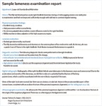
Sample lameness examination report
The first exam technique is close observation, Mitchell explains. The practitioner should be observant—putting his or her eyes and hands on the horse is essential. Detailed record keeping should document the lameness exam, which includes the following:
> Visual examination. Take notice of the horse's body symmetry and muscle structure.
> Stall observation. Observing the horse in its stall may reveal its comfort level, while observing the horse walk out of the stall may give clues about chronic lameness issues.
> Palpation examination. After the initial post-stall observation, a palpation examination should be performed and should include eliciting responses to potentially painful areas.
> Hoof examination. Flexion and hoof testers should be used prior to observing the horse in action.
> Working observation. Observe the horse in hand at a walk and trot in straight-line movement and in circles, followed by similar observation while jogging the horse on a lead. It is important to make these observations on soft and hard surfaces.
> Flexion tests. Perform flexion tests of all limbs while the horse is walking or trotting away.
> Wedge tests. After observing the horse in motion, wedge tests can be useful to localize the sites of lameness. "A reverse wedge that stretches the deep flexor tendon may increase a lameness related to that structure," Mitchell says. "A wedge placed laterally under the foot may stretch the medial collateral ligament of the distal interphalangeal joint and increase lameness if that structure is injured."1
Mitchell points out that practitioners should take care to not overexert a very lame horse until the nature of the lameness is recognized.
> Observation with rider. Once the horse is examined in hand, conduct similar observations with a rider present. Doing this may reveal distal limb pain or other problems that could be contributing to lameness, such as back or neck pain. The riding portion should include movement in straight lines and circles and on both soft and hard surfaces.
Other diagnostics
Once the practitioner has acquired additional information through the clinical examination, further diagnostics (e.g. nerve blocks) should be conducted. Depending on exam results, radiographs, nuclear scintigraphy, computed tomography (CT) or magnetic resonance imaging (MRI) may be employed if available and warranted.

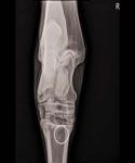
Imaging may be performed as part of a lameness examination but should never take the place of a thorough physical exam. In this case, a radiograph revealed a circular lytic lesion in the proximomedial aspect of the cannon bone (see sample lameness examination report for more details). (RADIOGRAPH COURTESY OF DR. HOGAN)
"A lot of young veterinarians might want to engage various technologies before they have established where the problem is," Mitchell says. "I do think imaging is important, but people often leap to imaging too quickly."
Mitchell states that if there's obvious pathology, such as a bowed tendon, a swollen leg that's hot and sore or a weight-bearing issue—injuries that are readily apparent—it's not necessary to employ nerve blocks. "Nothing replaces a thorough clinical examination," he says. "That cannot be emphasized enough."

Dr. Richard Mitchell flexes a horseâs front leg as part of a lameness examination. (PHOTO COURTESY OF DR. MITCHELL)
For the average practitioner who may not have advanced diagnostic equipment, Mitchell encourages a thorough workup to establish the location of the lameness and establish a case for referral to acquire specific imaging.
"Referring lameness cases and being responsible to the client can be a practice builder," he says. "We have built some wonderful relationships with referring veterinarians. We all help one another out, and we all benefit. When it comes to building one's business, the relationships between referring veterinarians and other practices can't be overemphasized."
Ed Kane, PhD, is a researcher and consultant in animal nutrition. He is an author and editor on nutrition, physiology and veterinary medicine with a background in horses, pets and livestock. Kane is based in Seattle.
Reference
1. Mitchell, R. Distal limb lameness in the sport horse: A clinical approach to diagnosis, in AAEP Conference Proceedings 2013;59:244.