Addressing the hidden defect in the leaky puppy
Young dogs have some simple reasons for leaving their urine where it doesn’t belong. But the not-so-simple ectopic ureter, a rare and often overlooked cause, can wreak havoc in the home.
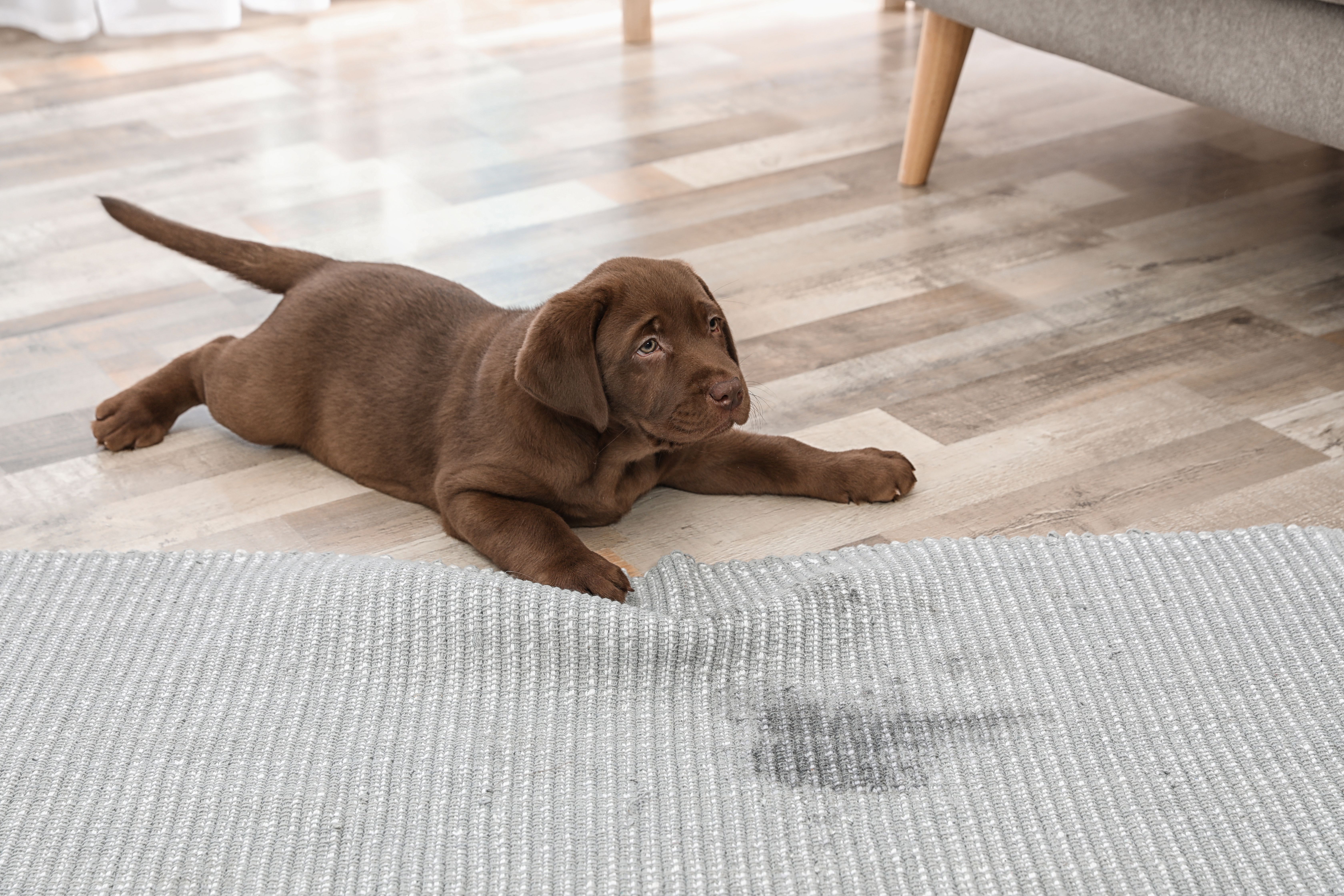
“My puppy is peeing all over the house!” We’ve all heard this distress call from panicked puppy owners. And we assure them that there is no reason for alarm. Housebreaking takes time. A puppy’s bladder muscles are not fully developed until age 4 to 6 months; a 10-week-old pup can only hold urine for a couple of hours. Of course there are going to be accidents around the house.
Normal plumbing
The urinary tract is a complex blood filtration system. It rids the body of waste and excess water. Blood purification starts out in 2 richly vascular kidneys, which retain what the body needs and discard everything else as urine. The urine exits each kidney through the ureter, a long, slender tube that attaches to the bladder. Here, the urine is stored for later voluntary release into the piped urethra and then out into the world.
The dripping faucet
The ureters—vital conduits for normal urinary evacuation—can be wrongly positioned because of an embryologic glitch in a pup’s development. Instead of terminating at either side of the bladder’s midsection, 1 or both ureters open into a different spot, like the urethra, vagina, or even uterus. Because the pump function of the balloon like bladder is disrupted, urine trickles out on a constant basis.
“It’s uncomfortable for a dog to be constantly leaking urine and wet all the time,” says Dana Clarke, VMD, DACVECC, assistant professor of interventional radiology at the University of Pennsylvania School of Veterinary Medicine, “and it’s incredibly frustrating for the owner, especially when it’s a big dog.”
Ectopic ureters, which are often accompanied by other urinary tract abnormalities, are rare overall. However, they are the most common congenital (birth defect) cause of canine urinary incontinence.
Clinical presentation
Although ectopic ureter is a problem that exists at birth, the obscurity of housebreaking usually delays diagnosis. Most affected pups do not land in the veterinarian’s office to address the issue until 6 to 12 months of age. Females compose 90% of cases, and affected males typically present closer to age 2.
Ectopic ureter has been reported in littermates, but the mode of inheritance is not fully understood. The breeds most commonly affected are Labrador and golden retrievers, West Highland white and fox terriers, English bulldogs, Siberian huskies, and Newfoundlands. Ectopic ureter occurs rarely in cats and horses, but has been reported in cattle, sheep, and pigs. Approximately 1 in 500 human babies are born with the condition.
Clinical signs in dogs include the following:
- Persistent wet hind end and bedding
- Regular urine dribbling, accompanied by episodes of normal urination
- Licking of the genital region, and urine scalding and rashes in the area
- Frequent bladder infections
Diagnosing ectopic ureter
Identifying ectopic ureter can be challenging because the condition is often mistaken for urinary tract infections or incomplete housebreaking. Routine testing, including blood work and urinalysis, is performed. Because urinary tract infections resulting from urine pooling in the urethra and vulva often accompany ectopic ureter, antibiotics are administered and sometimes provide mild resolution of signs. But for pups with ectopic ureter, the story just continues, as should the diagnostics.
Radiography and/or ultrasound can be performed to rule out bladder stones and anatomic abnormalities, but they do not display the thin ureters. To visualize these, an intravenous pyelogram—an x-ray that incorporates injected dye to visualize the ureters—or a CT scan is typically done.
These tests can be used not only to diagnose ectopic ureter but also to image damage secondary to the condition. Because the ectopic ureteral opening to the bladder or other organ is often blunted or partially blocked, sort of like a twisted garden hose, urine can back up and swell the ureter (hydroureter) and the delicate kidney (hydronephrosis) that lies upstream.
Once ectopic ureter is confirmed via imaging and the chaos it has created within the urinary tract is clarified, the precise pathway of the ureters and their opening point(s) are identified with cystoscopy. Here, a scope is inserted to examine the lower urinary tract, trace the pathway of the terminal segment of the ureter, spot the location of the ureteral orifice, and assess urine flow through the opening.
Redirecting urine flow
The only way to close the puppy’s flood gates is through either traditional or laser surgery. The approach depends on the type of ectopic ureter present.
Most canine ectopic ureters are intramural. Rather than attach cleanly to the bladder, an intramural ureter tunnels through the bladder wall and opens into the wrong portion of the bladder; the opening may be choked. An extramural ureter, on the other hand, may have a normal attachment, but it hooks onto the wrong organ, such as the urethra.
For an intramural ureter, a neoureterostomy can be performed. Here, the bladder is opened surgically and a new opening is made at the appropriate point in the wall-entrapped ureter. Any portion of ureter that extends beyond the bladder is excised.
For the preferred minimally invasive version of this procedure, a cystoscope-guided laser is inserted through the urethra and into the bladder to zap the ureter until its opening is well within the bladder, and to destroy any overhanging ureter.
“Endoscopy is the gold standard because it allows us to do both the diagnosis and the treatment in one procedure, with no restrictions for the dog afterward,” Clarke explains.
The extramural ureter is adjusted surgically by neoureterocystostomy. Here, the abdomen is opened and the ureter is detached from its abnormal location and reattached to the bladder.
Surgery doesn’t make every dog completely continent, and success rates are lower in females. After surgery, some dogs need lifelong medications to stay dry, such as phenylpropanolamine, which improves urethral sphincter muscle tone.
Conclusion
The ectopic ureter can take the new owner down a path they never imagined when acquiring that cute puppy. But the good news is that therapeutic measures are available to turn a flawed pup into a manageable pet.
Joan Capuzzi, VMD, is a small animal veterinarian and journalist based in the Philadelphia area.

2 Commerce Drive
Cranbury, NJ 08512
All rights reserved.
