Is surgical removal of the eye warranted?
Is surgical removal of the eye warranted?
Signalment:
Canine, Shih Tzu, 10 years old, male neutered, 20.7 lbs.
Clinical history:
The dog presents today for a problem with the right eye and potential blindness.
Physical examination:
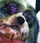
The findings include rectal temperature 102.2° F, heart rate 130/min, pink mucous membranes and normal capillary refill time. The dog is bright, alert and responsive. Normal heart and lung sounds are heard. Ocular examination shows corneal ulcer, buphthalmos and blindness in the right eye. A proliferative intraocular mass is extending through the globe of the right eye. Vision is normal in the left eye.

Table 1: Results of laboratory tests
Laboratory results:
A complete blood count, serum chemistry profile and urinalysis were performed and are detailed in Table 1.
Radiograph examination:
The thoracic radiographs are normal -- no obvious evidence of metastatic disease noted. The abdominal radiographs show an air-filled stomach, slightly enlarged liver, cystic calculi, enlarged prostate gland and severe osteoarthritis of the coxofemoral joints.

Image 1.
Ultrasound examination:
Thorough abdominal and right ocular ultrasonography was performed.
My comments:
The liver shows an inhomogeneous to homogeneous texture in its parenchyma. No masses noted within the liver parenchyma.
The gall bladder is mildly distended, and its walls are not thickened or hyperechoic.
The gall bladder does contain some sludge material. The common bile duct is slightly dilated. The spleen shows an inhomogeneous texture in its parenchyma - no masses noted. The left and right kidneys are similar in size, shape and echotexture. Each kidney shows an inhomogeneous texture in the renal cortex. No masses or calculi were noted in either kidney.

Image 2.
The urinary bladder is distended with urine and contains a lot of urine sediment material - no masses noted. There are multiple calculi present in the lumen of the urinary bladder and proximal urethra.
The prostate gland is slightly enlarged, symmetrical in shape, and possible calculus or consolidated sludge material present within the prostatic urethra. The left and right adrenal glands are similar in size and shape. The stomach, small intestines, and colon are normal. The pancreas shows an inhomogeneous texture in its parenchyma.
The right eye shows a large mixed echogenic, irregular-shaped, mass-like structure that occupies the anterior chamber. The posterior lens is not seen. Multiple, irregular-shaped, small echogenic structures are seen in the posterior chamber.

Image 3.
One or two round-shaped echogenic areas are caudal to the optic disk in the extraocular area surrounding the optic nerve. The left eye is normal in the anterior chamber, posterior chamber, posterior lens, and optic disk.
Case management:
In this case, right ocular mass and cystic calculi are the clinical diagnosis. The right ocular mass is most likely a melanogenic neoplasia. There was no obvious evidence of metastatic disease noted during this abdominal ultrasound study.
In addition, there is no obvious radiographic evidence of intrathoracic and intra-abdominal masses noted. At this point, surgical removal of the right eye and mass is warranted.

Image 4.
Therapy thereafter would depend on the findings from the histopathologic examination after right eye removal. Therapy for the cystic calculi could include feeding exclusively Waltham's S/O diet.
A bacterial urine culture is warranted and daily antibiotics administered according to the results of the urine culture. After about eight weeks of dietary management, cystic calculi may be resolved.
Ocular and orbital imaging technique
Imaging of ocular and orbital structures has been improved by the development of ultrasonography, computed tomography and magnetic resonance imaging (MRI).
Two-dimensional real-time ultrasonography of the eye and orbit is ideally suited for examination of opaque eyes and soft tissues of the retrobulbar space. It can usually be performed in awake and unsedated animals and provides good imaging detail of soft tissues.

Image 5.
Ultrasonograms may be acquired using an eyelid contact method, a corneal contact method or a gel offset method. The eyelid contact and gel offset methods allow better delineation of the cornea and anterior chamber. The direct corneal method is used when posterior segment or retrobulbar disease is suspected.
The ultrasonography may be performed with either sector scanners or linear array transducers, with 7.5 MHz or 10 MHz probes providing the best resolution. Imaging is useful in identifying intraocular lesions such as lens luxations, retinal detachments, hemorrhages, masses and certain foreign bodies, especially in eyes with an opaque cornea or lens. Color Doppler ultrasonography has some clinical usefulness. Color Doppler ultrasonography identifies the major vessels or vascular regions of the orbit and allows diseased orbits to be classified as hypo-, hyper-, or neovascularized.
Fact sheet
The melanoma is the most common intraocular tumor. The primary melanogenic tumor in dogs is the anterior uveal melanoma. The canine melanogenic mass is usually localized in the anterior uveal tract with low metastatic potential, whereas feline melanogenic mass is localized in the anterior uveal tract with high metastatic potential.

Image 6.
The clinical appearance of melanogenic neoplasia is heavily pigmented and tan to white or diffuse iris thickening. The melanogenic neoplasia causes an irregular pupil, blindness and ocular pain.
Glaucoma is secondary to the exfoliation or proliferation of cells within the uveal tract. Local infiltration and destruction occur as the neoplasm grows. Enucleation is usually required although laser surgery may be used to selectively reduce intraocular masses. In addition, a sector iridectomy is possible for uveal mass excision. Limbal and scleral melanocytic neoplasms (epibulbar melanoma) in dogs are usually benign.
The epibulbar melanoma often arises in the superior temporal quadrant of the eye and is slower growing, less invasive and well delineated in the sclera of older dogs. As the normal canine globe has melanocytes traversing the limbic sclera, conversion to abnormal growth occurs when the pigmented cells proliferate and invade the sclera, cornea and conjunctiva.
Surgical correction by full thickness corneal or scleral resection, laser reduction or cryosurgery may be curative since growth and extension of the neoplasm is slow.
Choroidal melanomas are infrequently seen in older dogs. Clinical signs include raised darkly pigmented areas of the fundus and domed thickening of the choroid with extension to the iris.
Dr. Hoskins is owner of DocuTech Services in Baton Rouge, La. He is a diplomate of the American College of Veterinary Internal Medicine with specialties in small animal pediatrics. He can be reached at (225) 955-3252; fax: (214) 242-2200.


