Uses and limitations of ECG, Doppler, and Capnograph
Consider these important factors when designing an operating room
Ekaterina / stock.adobe.com

If you are building or retooling your operating room, then you need to consider your anesthetic equipment. Proper tools and training are essential because poor anesthetic monitoring has consequences, such as worsening of chronic renal and cardiac diseases, gastrointestinal issues, the development of chronic pain, or even death. Minimum standards for anesthetic safety include continuous monitoring of respiratory function, circulation, heart rate, pulse, perfusion quality, oxygenation, ventilation, and temperature, which carry all the way through to recovery.
What follows are some of the most basic pieces of equipment a veterinary practice might have, along with their uses and limitations.
Electrocardiogram
Uses
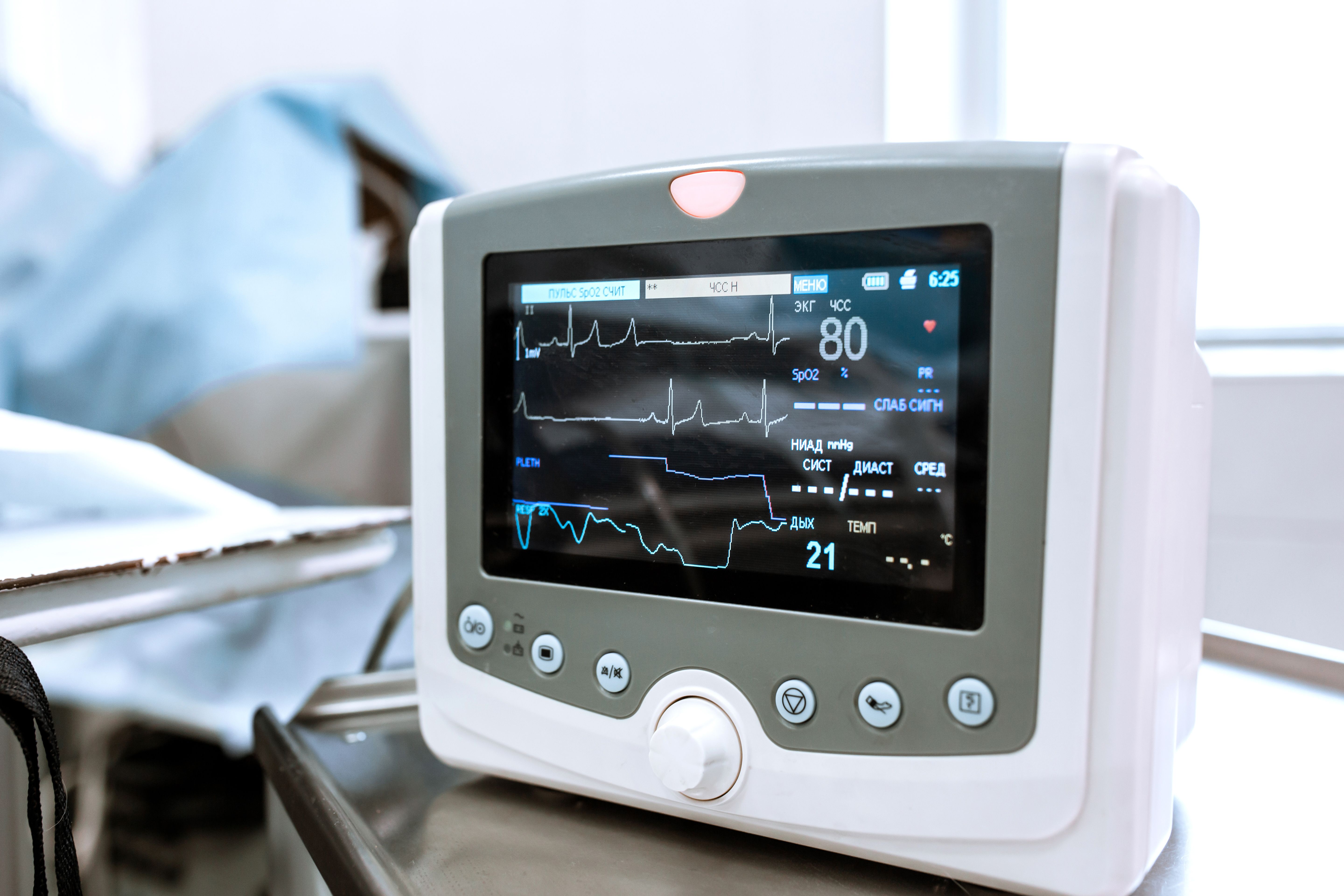
One of the most important pieces of monitoring equipment is the electrocardiogram (ECG). The function of the ECG is to provide real-time assessment of cardiac rate and rhythm and to signal changes in cardiac electrical patterns. General anesthesia may uncover underlying issues in patients that are not known beforehand. The ECG is also essential to screen patients for extracardiac issues such as electrolyte abnormalities and hypoxemia. Most ECGs are now combined into multiparameter monitors with several different systems conveniently in the same unit.
Limitations
Artifacts may be caused by electrical interference from cautery, nerve stimulators, other heating devices, and more. Your team must be skilled enough to determine whether something is an artifact or a real issue. For example, pulseless electrical activity—characterized by a complete dissociation between the cardiac electrical activity and its contractility—is one of the most severe issues and can manifest as a slightly abnormal ECG, despite the fact that the animal has actually gone into cardiac arrest.
Doppler Flow Detector
Uses
Another means of monitoring circulation and pulse is a Doppler flow detector, a unit that converts blood flow into an audible signal. The portable unit can travel with your patient from preop to recovery and is a noninvasive and reliable way to monitor continuous pulse and heart rate. It provides an opportunity to detect any infrequent or subtle arrhythmias.
Limitations
—Certain classes of drugs may affect the sound quality of the pulse.
—Attaching a probe to the patient is an art form because it must be securely attached—not too loose, not too tight.
—The patient needs to remain reasonably motionless.
—The unit may pick up radiofrequency interference that distorts the sound.
—Any damage to the crystals will also affect sound quality.
—You cannot determine whether an arrhythmia you hear audibly is sinus or ventricular in origin.
Capnography
Uses
Capnography is one of the most important tools in anesthesiology. Ventilation monitoring is primarily through measurement of end-tidal carbon dioxide (CO2), done through a noninvasive attachment placed onto the patient’s endotracheal tube that provides confirmation of correct placement in the trachea and adequacy of patient ventilation.End-tidal CO2 is also used as a marker for cardiopulmonary resuscitation effectiveness. The wave form produced by a capnogram also detects any disconnection or obstruction and warns of impending cardiac arrest.
Two types of CO2 sensors are available: mainstream and side stream. Mainstream units measure directly at the sample site, which may be bulky for small patients but has no delay in measurement. A side-stream sensor draws gas from the patient and measures it at a distant site, usually at the base of the monitor.
Limitations
—Lines can become kinked or obstructed by water or secretions, causing issues with the waveform.
—Humidification, tiny cracks or gaps in the connections, and loose connection can cause artifacts.
Closing words
There are many factors to weigh regarding anesthetic equipment in the operating room. Careful consideration on the uses and limitations of each item can play a major role in maintaining a high level of quality and care in anesthetic monitoring.
