What are the medical problems with intra-abdominal neoplasia?
Surgery is the only meaningful option for this cat.
Editorâs Note: In our ongoing telemedicine series, Dr. Johnny Hoskins presents medical case studies. The format is focused heavily on radiology and ultrasonography and details complicated yet comprises fairly common cases that most veterinarians will be exposed to in practice.
Signalment:
Feline, Persian, 10 years 9 months old male neutered, 7.5 lbs.

Figures 1-7 (Click to enlarge)
Clinical history:
The cat presented for sniffling and eating well but not as much as normal. The cat is FIP positive. Owner suspected URI.
Physical examination:
The findings show rectal temperature of 101.8°F, heart rate 180 beats/min, sinus rhythm, respiration 20 breaths/min, pink mucous membranes and no heart murmur heard. Soft tissue mass is palpated in the middle to cranial abdomen.
Laboratory results:
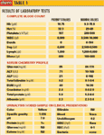
A complete blood count, serum chemistry profile and urinalysis were performed and are outlined in Table 1.
Radiographic review:
Survey thoracic and abdominal radiographs are done. The thoracic radiographic views are unremarkable. The abdominal radiographs are depicted in Images 1 and 2 (p. 20S).
My comments:
The abdominal radiographs show a mass effect in the mid to cranial abdomen and a relative lack of accumulated intra-abdominal fat.
Ultrasound examination:
Thorough abdominal ultrasound is performed with the cat positioned in dorsal recumbency. (See Images 3 through 7.)
My comments:
The liver shows a uniform echogenicity in its parenchyma. No masses noted within the liver parenchyma. The gall bladder is mildly distended, and its walls are not thickened or hyperechoic. The gall bladder does contain some sludge material. The spleen shows a uniform echogenicity in its parenchyma — no masses noted. The left and right kidneys are similar in size, shape and echotexture. Each kidney has cortical cysts. No masses or calculi were noted in either kidney.

Image 8
The urinary bladder is distended with urine and contains some urine sediment material — no masses or calculi noted. The stomach is normal.
Case management:
The tentative diagnosis is mild renal disease and intra-abdominal neoplasia. I am not sure of the origin of this cat's neoplastic process. Surgery is the only meaningful option for this cat. At this point, an exploratory laparotomy would be warranted to attempt removal of the neoplastic process, inspect the abdominal cavity for potential metastatic disease, and collect appropriate biopsies for histopathologic examination.

Image 9
Therapy thereafter would depend on the findings from the exploratory laparotomy and histopathologic examination.
Surgical management:
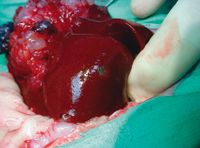
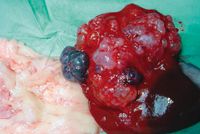
Exploratory surgery was done. See Images 8-13 (p. 22S and this page).

Table 1: Results of laboratory tests
These are photos of the mass on the liver and cystic masses throughout the small intestines. Section of tissue from the liver consists of a tumor mass. The tumor consists of epithelial cells forming numerous large cystic structures. Some of these cysts are filled with blood. The lining of the cyst is composed of flattened cuboidal epithelial cells. Some remnant liver is present in between cysts. Histopathologic results: Biliary Cystadenoma.

AKC awards $160,000 in veterinary scholarships