ACVP 2017: Overview of Ferret Lymphoma
Practitioners who treat small mammals should understand the clinical variants, presentation, and staging and classification of this prevalent disease.

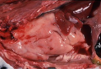
Thymic lymphoma in a ferret.
Lymphoma is common in ferrets, according to Bruce H. Williams, DVM, DACVP, senior pathologist at the Joint Pathology Center in Silver Spring, Maryland. “Lymphoma is the third most common tumor overall in ferrets and by far the most common malignancy,” he said.
In his presentation at the 2017 annual meeting of the American College of Veterinary Pathologists (ACVP) in Vancouver, British Columbia, Canada, Dr. Williams reviewed some important aspects of the different variants of lymphoma in ferrets. There is no gender predilection for lymphoma in ferrets, he said, although—as is the case with most neoplasms—its prevalence tends to increase with age. The “golden years” for tumor development in ferrets are between ages 4.5 and 9 years, and especially regarding the endocrine and hematopoietic systems, Dr. Williams said. He explained that over the past 10 or 20 years, the average life span of ferrets has increased from about 7 years to 9 years due to better understanding of these animals. However, he added, a longer life span also increases opportunities for tumor development.
RELATED:
- FDA Conditionally Approves Tanovea-CA1, the First New Animal Drug for Treating Lymphoma in Dogs
- Feline Lymphoma: How Has It Changed?
Although lymphoma typically arises spontaneously in ferrets, some studies have suggested that it may also be transmissible, prompting speculation of an association with a type C retroviral infection.1 Nevertheless, research in this area is lacking after identification of retroviral particles almost 20 years ago,1 and this association remains unproven.
Sites of Origin of Lymphoma in Ferrets
“There is no typical picture for lymphoma in the ferret,” Dr. Williams said. “It may pop up in any organ, and the associated clinical picture usually reflects dysfunction of that particular organ.” However, several variants of lymphoma, based on site of origin, have been reported.
Multicentric Lymphoma
The most common form, multicentric lymphoma typically arises in older ferrets. It presents as peripheral lymphadenopathy and spreads to the spleen and other visceral organs, leading to organ failure late in the course of disease.
Mediastinal Lymphoma
This is the second most common form and occurs mostly in younger ferrets, typically those under age 2 years. It characteristically involves the thymus and the lymph nodes in the thorax, but the disease may also spread to involve other viscera.
Gastrointestinal Lymphoma
Gastrointestinal (GI) lymphoma is a relatively uncommon form of this disease, although it is the most common malignancy occurring in the GI tract. GI lymphoma has the shortest survival time of the variants; affected animals typically survive only about 2 weeks after presentation. It potentially arises from chronic inflammation in the GI tract and may be associated with inflammatory bowel disease (IBD) or infection with organisms such as Helicobacter or coronavirus.
Helicobacter infection often occurs in ferrets after about 3 years of age. All infected animals tend to have associated GI inflammation, Dr. Williams said, although not all develop clinical signs of disease. Nevertheless, the inflammation tends to “set up” the GI tract to develop lymphoma, he explained.
Today, IBD is the most common diagnosis from GI biopsy submissions from ferrets, Dr. Williams said, adding that—at least from the pathologist’s viewpoint—GI lymphoma forms a spectrum of morphology with severe IBD. Although the exact etiology of IBD in ferrets remains unknown, “it also produces a fertile ground for expansion of clonal lymphocytes to then set up lymphoma,” he added.
For veterinarians managing ferrets with clinical signs suggestive of IBD and/or lymphoma, Dr. Williams recommended collecting full-thickness biopsy sections of the GI tract for pathologic evaluation. Most cases of GI lymphoma tend to be T-cell type, he said, followed by B-cell type and, distantly, null-cell types; however, studies have shown that immunohistochemistry is usually not required for differentiating between lymphoma and IBD.2
Cutaneous Lymphoma
Cutaneous lymphoma is uncommon in ferrets, existing in 1 of 2 forms. Epitheliotropic T-cell lymphoma is nonlethal, tends to affect older ferrets, and typically responds well to surgical removal if amenable. “Swollen feet can be 1 manifestation of epitheliotropic T-cell lymphoma,” Dr. Williams noted. In contrast, nonepitheliotropic lymphoma tends to be lethal and progressive and may occur as part of multicentric lymphoma. Skeletal Lymphoma
This form is also uncommon, with just single cases mentioned in the veterinary literature. Reports have described lymphoma arising in different regions of the skeleton and having different morphologies, including T-cell lymphoma in the mandible, humerus, and femur; plasmablastic lymphoma in a femur; and null-cell lymphoma in the thoracolumbar spine. Skeletal lymphomas tend to be very osteolytic, Dr. Williams said, and may extend into surrounding skeletal muscle.
Hodgkin-like Lymphoma
Another uncommon variant in ferrets—reported just 3 times—is Hodgkin-like lymphoma, involving either single lymph nodes or a chain of regional lymph nodes. Although these cases do not exactly match the criteria of Hodgkin lymphoma in humans, they are based on identification of characteristic Reed-Sternberg cells.
Common Presentations of Lymphoma in Ferrets
Lymphoma typically shows up in ferrets younger than 2 to 3 years of age and classically presents as visceral masses, Dr. Williams said. “Lymphoma in young ferrets tends to present very stereotypically,” he said. “They often have a large thymic mass and, while the peripheral lymph nodes remain untouched, the lymphoma may also be in other viscera such as the spleen.” The disease may thus present as acute-onset respiratory distress because of compression of the lung lobes, dyspnea, and pleural effusion. “Next to cardiovascular disease, lymphoma is one of the most common causes of effusions in ferrets,” Dr. Williams said. Young ferrets with lymphoma typically also have lymphocytosis.
Although this form of lymphoma tends to respond to prednisone for about 1 month, it soon recurs and is associated with a short survival time. Histologically, these cells tend to have a large-cell morphology, often being more than twice the size of a red blood cell. In contrast, lymphoma in older ferrets typically presents as peripheral lymphadenopathy—rather than visceral involvement, as in younger animals—and is characterized by a small-cell morphology (neoplastic cells are about the same size as normal lymphocytes). Affected animals typically have lymphopenia but tend to survive longer than younger ferrets with lymphoma do. These older ferrets survive up to 2 years, with or without chemotherapy, Dr. Williams said.
Because most cases of lymphoma in lymph nodes in ferrets can be identified on cytologic examination, Dr. Williams shared some pointers for veterinarians who wish to perform fine-needle aspiration biopsy of enlarged lymph nodes. “If it’s not big, don’t aspirate it,” he emphasized.
Importantly, he also reminded the audience of the difference between normal and neoplastic lymph nodes. Because of surrounding fat, peripheral lymph nodes in older ferrets tend to feel relatively soft. In contrast, “neoplastic lymph nodes tend to be very hard to the touch,” Dr. Williams said. He also advised veterinarians to preferentially sample the popliteal node rather than the mandibular or mesenteric node.
Although cytology can be used to diagnose lymphoma in enlarged lymph nodes in ferrets, he reminded veterinarians that staging and classifying lesions requires microscopic evaluation of excised nodes.
Lymphoma Staging and Classification
There is no universal staging system specifically for lymphoma in ferrets, but Dr. Williams said that the 5-level clinical staging definitions developed by the World Health Organization (WHO) can be used (Table).
In ferrets, most cases of lymphoma tend to be stage IV; certainly, lymphoma in young ferrets typically presents as stage IV, diffuse, high-grade, large-cell lymphoma, Dr. Williams said. In older ferrets, it also often presents as stage IV, diffuse, high-grade, small-cell lymphoma but also as stage II, diffuse, low-grade, small-cell lymphoma. Another exception, he noted, is Hodgkin-like lymphoma, which falls into the stage I category.
The results of a recent study showed that veterinary pathologists can use an adaptation of the latest WHO classification for neoplastic diseases of the lymphoid tissues to diagnose lymphomas in dogs accurately.3 This adaptation grades lesions based on criteria such as the size of neoplastic cells and prevalence of mitoses.
To improve understanding of ferret lymphoma, Dr. Williams suggested that veterinary pathologists consistently apply these standardized criteria when evaluating these tumors. That will allow everyone “to begin speaking the same language,” he concluded.
Dr. Parry, a board-certified veterinary pathologist, graduated from the University of Liverpool in 1997. After 13 years in academia, she founded Midwest Veterinary Pathology, LLC, where she now works as a private consultant. Dr. Parry writes regularly for veterinary organizations and publications.
References:
- Erdman SE, Reimann KA, Moore FM, Kanki PJ, Yu QC, Fox JG. Transmission of a chronic lymphoproliferative syndrome in ferrets. Lab Invest. 1995;72(5):539-546.
- Watson MK, Cazzini P, Mayer J, et al. Histology and immunohistochemistry of severe inflammatory bowel disease versus lymphoma in the ferret (Mustela putorius furo). J Vet Diagn Invest. 2016;28(3):198-206. doi: 10.1177/1040638716641156
- Valli VE, San Myint M, Berthel A, et al. Classification of canine malignant lymphomas according to the World Health Organization criteria. Vet Pathol. 2011;48(1):198-211. doi: 10.1177/0300985810379428
