ACVC 2017: Understanding Skin Disease in Cats
Although much remains unknown about feline dermatology, one thing is certain: Many skin conditions are related to internal disease.

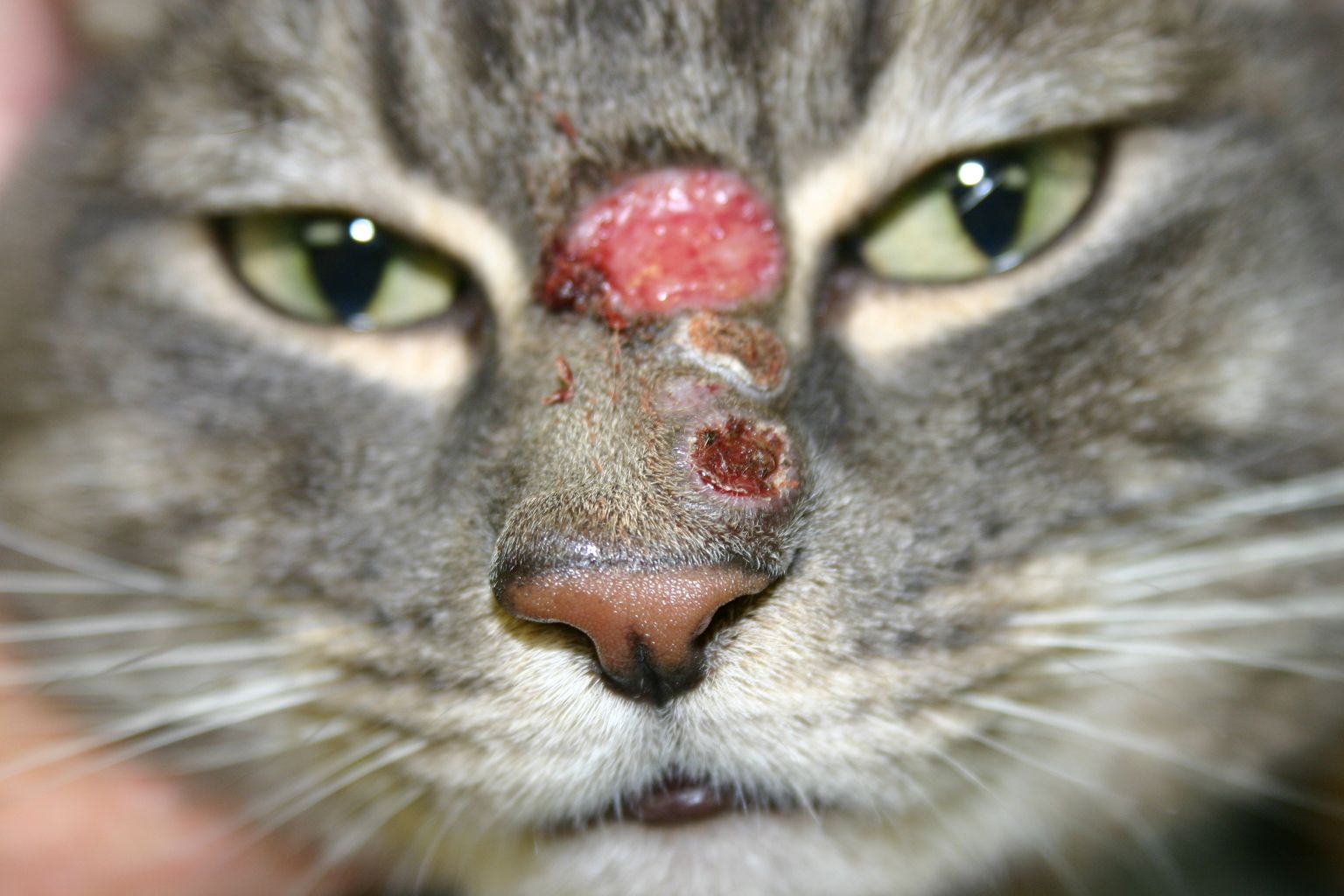
Facial herpes.
When it comes to skin disease, cats are not just small dogs. This was a key point from a presentation on skin disease in cats by Candace A. Sousa, DVM, DABVP (Emeritus; Canine & Feline Practice), DACVD, at the 2017 Atlantic Coast Veterinary Conference in Atlantic City, New Jersey. “Cats are cats,” she said, “and their disease processes need to be approached from a cat point of view.”
Feline skin diseases may involve a severe underlying illness and require a thorough diagnostic evaluation, including a detailed history and physical examination. Getting a diagnosis generally makes therapy and prognosis “easy,” Dr. Sousa said.
Unlike internal medicine, veterinary dermatology has limited diagnostic tests (eg, skin cytology, trichogram, biopsy and analytic tools (eg, Wood’s lamp, microscope) available; trichograms, in particular, are performed more often in cats than in dogs. Veterinarians should charge for each skin diagnostic test, including simple scrapings, Dr. Sousa said, emphasizing that clients should pay for the expert opinion and knowledge. In addition to obtaining a history and performing a physical exam, organizing possible skin diseases into categories can help in making a diagnosis. Categories include allergic, bacterial, congenital/ hereditary, endocrine, fungal, immune-mediated, miscellaneous, neoplastic, nutritional, parasitic, and viral diseases.
RELATED:
- A Genetic Mutation Causes a Rare Skin Disease in Dogs
- New Insights on Feline Skin Microbiota
After making the diagnosis, Dr. Sousa recommended that veterinarians ask owners what they want to do regarding treatment—“What would you like?” or “What are your goals for your cat?”—being careful to avoid a paternalistic tone. She then provided details on congenital, endocrine, neoplastic, parasitic, viral, and miscellaneous skin diseases that are unique to cats.
Congenital Diseases
Idiopathic Facial Dermatitis of Persian Cats
Also known as “dirty face syndrome,” this chronic disease starts when Persian cats are young and causes black crusts on the face, particularly in the folds and periocular area. Affected cats may traumatize themselves. In addition, ceruminous otitis may be present concurrently. Treatment response is typically poor.
Proliferative and Necrotizing Otitis
Proliferative and necrotizing otitis is rare, has an unknown etiology, and typically affects 3- to 6-month-old kittens. Physical examination of the ears reveals large tan or dark coalescing plaques on the concave surface of the pinnae and external ear canals; the ears may also contain comedones. Histopathologic findings include acanthosis, follicular keratosis, and hair follicle outer sheath hyperplasia and keratinocyte necrosis.
This skin condition can be treated with topical 0.1% tacrolimus, which is expensive. Oral prednisolone can also be used, but its effectiveness in treating this disease remains unknown. The prognosis is poor.
Endocrine Diseases
Feline Skin Fragility
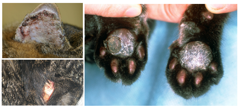
Typically a marker of feline hyperadrenocorticism, feline skin fragility generally occurs in older cats. With several potential causes (eg, progestin therapy, excessive administration of corticosteroids), this condition results in a collagen deficiency that makes the skin extremely thin.
Examining an affected cat requires very gentle handling to avoid tearing the skin. Interestingly, Dr. Sousa noted, not much bleeding occurs when the skin tears. Also, unlike in dogs, alopecia is typically not present in affected cats. Biopsies, although not practical because of the skin’s thinness and fragility, often show collagen deficiency and severe epidermal, dermal, and follicular atrophy. Dr. Sousa recommended that adrenal function be tested during the diagnostic evaluation. The prognosis is grave, especially if the underlying cause cannot be identified.
Neoplastic Diseases
Paraneoplastic Alopecia Associated With Visceral Neoplasia
This skin disease has an acute onset and generally affects cats aged 10 years and older. There is complete alopecia of the ven- trum; the skin appears shiny, potentially due to stratum corneum exfoliation. Scaly footpads are a hallmark of this disease.
Clinical signs (weight loss, anorexia, and lethargy) suggest an underlying systemic disease. Abdominal ultrasound may reveal visceral neoplasia, which is usually a pancreatic adenocarcinoma. Skin biopsy findings—severe follicular and adnexal atrophy; alopecia-associated epidermal thickening—can also aid in tumor identification. It is thought that the tumor releases factors, such as cytokines, that cause follicular shrinkage. The prognosis is grave.
Exfoliative Dermatitis and Thymoma Exfoliative Dermatitis
This rare form of dermatitis, secondary to an underlying thymoma, causes scaly and erythematous dermatitis that begins on the head and neck and eventually becomes generalized. The dermatitis is nonpruritic and affects middle-aged and older cats. It is thought that defective thymic lymphocyte selection results in an abnormal immune response to keratinocyte antigens, causing keratinocyte apoptosis.

Top left: Bowen disease on the inner right pinna. Bottom left: Skin fragility. Right: Plasma cell pododermatitis.
Histopathology findings include cell-poor interface dermatitis (dermatitis at the dermo-epidermal junction), mild hydropic degeneration of basal cells, and keratinocyte apoptosis. This feline skin disease is treatable with surgical resection of the tumor.
Metastatic Bronchogenic Adenocarcinoma
Older cats with lung tumors may develop this condition, also known as “lung digit.” Amazingly, Dr. Sousa said, affected cats typically show no signs of respiratory disease before skin abnormalities appear. The tumor metastasizes to the capillaries in the distal extremities and toes, causing an acute onset of hemorrhage in the digits. Without confirmation of a lung tumor, this skin disease may be mistaken for inflammatory pododermatitis.
Topical or systemic corticosteroids can be used to relieve edema and general discomfort, but the prognosis is poor.
Parasitic Diseases
Mosquito Bite Hypersensitivity
This seasonal disease is uncommon in cats but worth knowing about. Mosquitoes may bite thinly haired areas like the bridge of the nasal planum, the pinnae, and footpads, creating immediate and late-phase hypersensitivity reactions to the mosquito salivary antigens. Typically, the initial bite will develop into a wheal within 20 minutes; papules appear in 24 hours and become encrusted within about 48 hours.
Cats that are chronically affected by mosquito bite hypersensitivity develop lesions such as scaling, alopecia, and pigment changes. Histopathology, which is needed to confirm this skin disease, demonstrates highly eosinophilic dermatitis. Cats that become hypersensitive to mosquito bites are usually affected repeatedly. However, Dr. Sousa noted, not all cats that get bitten by mosquitoes develop a hypersensitivity reaction.
Management options include confining the cat in a mosquito-free environment and using topical mosquito repellent on areas susceptible to bites. Systemic corticosteroid treatment is indicated for cats that cannot be confined.
Viral Diseases
Feline Immunodeficiency Virus—Related Dermatitis
Cats with either feline immunodeficiency virus (FIV) or feline leukemia virus may get skin disease. FIV-related skin problems include abscesses, skin and ear bacterial infections, and mycotic infections. Some cats with FIV develop nonpruritic, generalized, papulocrustous lesions with concurrent alopecia and scaling, which are most severe on the head and limbs. On histopathology, FIV-related dermatitis demonstrates hydropic interface dermatitis and giant keratinocytes.
To date, treatment options for this skin disease have not been successful. Cutaneous Herpes Virus Infections
This condition typically affects older cats (8 to 9 years old) and can cause ulcerative and necrotizing facial dermatitis. The nasal planum, bridge of the nose, and periocular skin are commonly affected. Histopathologic findings include ulcerated epithelium, intranuclear inclusion bodies in keratinocytes (indicating herpes virus infection), multinucleated giant cells and keratinocytes in the surface and follicular epithelium, and interstitial mixed inflammatory dermatitis with eosinophils.
Systemic antibiotics are indicated to treat secondary bacterial infections, and acyclovir can help treat the viral infection. Corticosteroids can activate the virus and thus are not recommended for treating cutaneous herpes virus infections. Lysine has been proposed as a treatment option but, according to Dr. Sousa, hasn’t shown much efficacy. Human alpha-interferon is another option for treating cutaneous herpes virus infections, but cats eventually develop antibodies to it, making it less effective over time. None of the treatment options will get rid of the herpes virus carrier site within an affected cat’s body, Dr. Sousa said.
Multicentric Squamous Cell Carcinoma In Situ (Bowen Disease)
Bowen disease affects cats older than age 10 years and is triggered by papillomavirus. This slowly progressive disease causes eroded crusted papules and plaques that are frequently found on the head and neck but can also appear on the shoulders and forelegs. This disease is not solar induced and does not metastasize.
To obtain a diagnosis, Dr. Sousa recommended taking a 6-mm punch biopsy of the dermis and epidermis; epidermal abnormalities, she warned, may prevent proper skin healing. To remove the sting of lidocaine, she advised adding sodium bicarbonate (0.9 mL lidocaine, 0.1 mL bicarbonate) to the local anesthetic. She also suggested prerinsing the syringe with epinephrine to reduce bleeding. Epidermal dysplasia without basement membrane destruction is apparent on histopathology.
Surgical laser excision is the preferred treatment, with radiation and systemic retinoids recommended as adjunctive therapies. Vitamin A therapy may be useful for keratinocyte proliferation.
Miscellaneous Diseases
Indolent Ulcer (“Rodent Ulcer”)

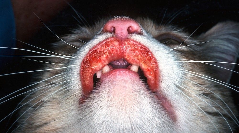
Indolent ulcer.
This skin disease has no age, sex, or breed predilection. Well-circumscribed, red-brown ulcers with raised borders usually form on the upper lip. Interestingly, ulcers are neither eosinophilic nor granulomatous on histopathology; peripheral eosinophilia may be present.
Chronic, excessive licking due to allergic or pruritic stimuli is thought to cause the ulcers. If the ulcers are recurrent or refractory, Dr. Sousa recommended testing for food allergies or flea allergy dermatitis.
Early treatment is best; however, systemic glucocorticoids are often ineffective. Systemic antibiotics and progestational drugs can be useful in refractory cases. Treatments such as cryosurgery and laser therapy are primarily cosmetic, Dr. Sousa said.
Eosinophilic Granuloma
The clinical description of this lesion varies with location. Eosinophilic granulomas are ulcerated on the lips or within the oral cavity (back of palate, palatal arches); on the caudal thigh, the granulomas are linear, raised, circumscribed, and yellow-to-pink plaques. On histopathology, eosinophils are attached to the collagen.
Linear granulomas can regress spontaneously. Granulomas on the lips or within the oral cavity can be treated with steroids.
Eosinophilic Plaques
This common feline skin condition also is not associated with a particular age, sex, or breed. Affected cats are extremely pruritic and resort to excessive licking and grooming, causing plaques. Most often there is an underlying allergic skin disease. The ulcerated lesions are typically found on the abdomen and medial thighs and are well circumscribed, raised, round or oval, and erythematous. Many cats have a circulating peripheral eosinophilia.
Treatment is the same as for indolent ulcers.
Plasma Cell Pododermatitis
This rare condition has no known etiology. Initially, the lesions, which frequently affect the central carpal or tarsal footpads, are soft and painless with loss of the surface architecture. Over time, the pads become ulcerated and develop secondary infections, causing pain, lameness, and regional lymphadenopathy. Affected cats are otherwise healthy.
Histopathology provides a definitive diagnosis. Spontaneous regression occurs occasionally, while other cases benefit from systemic glucocorticoid or prednisolone treatment; response begins within about 3 weeks and peaks within about 3 months. Tetracycline or doxycycline (5 mg/kg twice daily) can also be used. Surgical excision is recommended for ulcerated footpads to improve healing and prevent recurrence.
Injection-Site Panniculitis
Subcutaneous injections of rabies vaccine, praziquantel, and methylprednisolone acetate have reportedly caused panniculitis in the dorsal cervical region and midscapular area. Occurring several months after injection, the skin reaction can range from inflammation and alopecia to ulceration and fat necrosis.
Disease management can be difficult. Treatment options include intralesional or systemic glucocorticoids, identification and treatment of underlying allergies, and surgical excision.
Self-Induced Alopecia (“Fur Mowing”)
Affected cats often present with partial, symmetric, or complete alopecia on areas accessible to licking. Excessive licking may be due to a psychological disorder or underlying allergy. Trichograms of the distal hair tips will help demonstrate that the alopecia is self-induced.
If allergic and inflammatory conditions have been ruled out, medical treatment options—all oral—include phenobarbital (1-2 mg/kg once or twice daily), amitriptyline (5-10 mg/day), and buspirone (0.5 mg/kg/day), which may be also useful for psychogenic alopecia.
Dr. Pendergrass received her DVM from the Virginia-Maryland College of Veterinary Medicine and completed a postdoctoral fellowship at Emory University’s Yerkes National Primate Research Center. She is the founder and owner of JPen Communications, LLC, a medical communications company.
