


Case studies will be utilized to highlight major points in this presentation.
Cardiology
Latest News
Advertisement
CME Content
Advertisement
Electrocardiography is an integral part of the cardiological exam. It is the only way to determine heart rhythm accurately and to determine if there are any conduction abnormalities.
CDVD is the most common cause of cardiac disability in dogs. The disease process is best described as myxomatous degeneration of the heart valves wherein the integrity of the valves is compromised often resulting in valvular insufficiencies.
Diuretics (reduction in preload), vasodilators (reduction in preload or afterload), angiotensin converting-enzyme inhibitors (reduce afterload and preload, reduce fibrosis), and positive inotropic drugs (increase contractility, may reduce regurgitant volume) all have demonstrated the capacity to lessen the severity of mitral regurgitation and dilated cardiomyopathy under certain conditions.
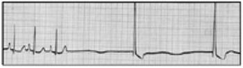
The electrocardiogram (ECG) is a commonly employed diagnostic tool to help in the evaluation of cardiac arrhythmias, to help detect cardiac chamber enlargement, and to identify electrolyte abnormalities.
A systematic approach to the evaluation of the ECG will ensure against overlooking important abnormalities.
The cause(s) of dilated cardiomyopathy (DCM) in dogs is (are) unknown. Some of the proposed causes of DCM include: genetic defect(s), viral infection, microvascular spasm, chemical toxin(s), dietary deficiency, and immune-mediated processes.
The primary objectives of the cardiovascular evaluation for animals with congenital heart disease are to define the nature and severity of the anatomic defect present. Familiarity with the available therapeutic options, their efficacy and limitations is necessary before an accurate prognosis can be offered to the owner.
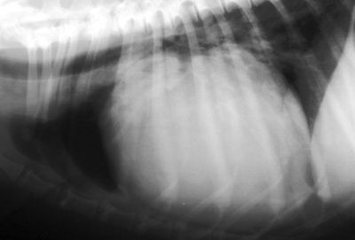
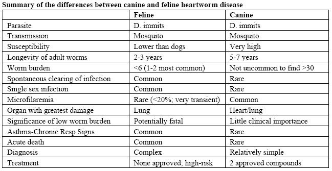
Myocardial disease is the most frequently diagnosed type of heart disease in the cat.
Read the ACVIM's consensus statement "Guidelines for the Diagnosis and Treatment of Canine Chronic Valvular Heart Disease."
These guidelines provide a framework for veterinarians to think about diagnostic strategies to screen for the presence of valvular disease.
To more objectively categorize patients and to complement the current classification system, the American College of Veterinary Internal Medicine (ACVIM) Specialty of Cardiology consensus panel has developed a set of guidelines for the diagnosis and treatment of CVHD.

A Q&A with veterinary cardiologist Stephen J. Ettinger, DVM
Echocardiography has emerged as the most valuable non-invasive tool for evaluation of cardiac structure, function, blood flow patterns, and has greatly diminished the need for diagnostic cardiac catheterizations and angiocardiography in many cases.
The primary indication for obtaining an electrocardiogram (ECG) is to evaluate an arrhythmia.
Feline heart disease is common and can be challenging to diagnose.
Congestive heart failure (CHF) is a common sequela to severe cardiac disease.
Pericardial effusion is a fairly common acquired heart disease in dogs, and prevalence has been reported to be 0.43% (or 1 dog per 233 cases) of dogs presenting to a referral veterinary hospital, and accounts for approximately 7% of dogs with clinical signs of cardiac disease.
Tips for improving cardiology diagnostics at your veterinary practice.
Your daily dose of clinical tips and quips: Cardiovascular exam.
Free heart health screenings give dogs a place to heal on four wheels.
Let's look at the applicability of measuring blood pressure, methods of assessment and the interpretation of results in clinical practice.
Predisposing factors that may preclude a cardiocerebral arrest should be eliminated when possible for every critical patient.
Successful CPCR is dependent on several factors, the most important factor being the true cause of the arrest.
Advertisement
Advertisement
Trending on dvm360
1
Q&A: Managing anemia, appetite loss, and phosphorus in feline chronic kidney disease
2
How Do I Say...?: Preblocked scheduling that ends daily chaos
3
Applying the principles of intravenous drug delivery
4
Keeping senior pets hydrated and comfortable all summer long
5