Study links bone loss to proximal sesamoid fractures in racehorses
Results of this retrospective study of California racehorses may mean that one day this type of fracture can be prevented.
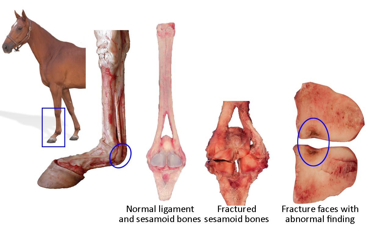
Sesamoid bone location, anatomy, and fracture. [Credit: Susan Stover, DVM, PhD, DACVS]

The proximal sesamoid bones (PSBs) are 2 relatively small bones located in the suspensory apparatus of the fetlock. The most proximal portion of the PSBs acts as the point of insertion for the suspensory ligament. The base of the PSBs acts as the point of origin for the distal sesamoidean ligaments.
PSB fractures are the most common fatal injury in racehorses, accounting for 45% to 50% of such injuries in racing Thoroughbreds and 37% to 40% in racing Quarter Horses.1,2 Although these fractures are seen in younger racing horses, they are more common in 3- to 5-year-old horses that have done a lot of training and racing.
Looking for a cause
In a retrospective study recently published in Equine Veterinary Journal, a group of researchers from the University of California, Davis, School of Veterinary Medicine characterized bone abnormalities that precede PSB fractures.3 The study consisted of 2 independent studies, each comparing the findings of 3 groups of PSBs:
- Fractured medial PSBs from racehorses that sustained PSB fracture
- The contralateral intact medial PSB from racehorses that sustained PSB fracture
- Intact medial PSBs from control racehorses that died from a cause unrelated to PSB fracture
Study 1 examined morphologic findings and serial sagittal sections from whole PSBs. Study 2 compared tissue morphometric properties from microcomputed tomography scans.
According to previous studies,4,5 PSB fractures result from chronic repetitive loading. Racehorses at higher risk for PSB fractures are those in active intensive training and racing, competing in more events at high speed/greater distance. “Understanding the mechanism of PSB fracture is necessary to reduce fracture risk and determine risk factors and fracture prevention,” the authors of the current study wrote.3
Study findings
The investigators noted abnormal findings in 2 specific locations within the PSB: the subchondral bone on the abaxial half of the PSB and (less commonly) along the palmar surface. They found that 90% of the injured horses showed discoloration at a site of focal osteopenia (bone loss) associated with the PSB fracture. A similar but less severe abnormality was commonly found in the contralateral, intact PSB of horses with PSB fracture, but this was not found in the control horses that did not have a PSB fracture.
To determine whether pre-existing conditions are associated with these fractures, PSBs were examined from Thoroughbred racehorses that died from either PSB or non-PSB fractures. Those horses that died from PSB fractures had evidence of abnormalities on the subchondral surface, including focal surface discoloration, focal osteopenia, subchondral bone sclerosis, focal fragmentation of subchondral bone, and calcified cartilage matrix.3 Subchondral lesions seen in the medial PSBs of racehorses may have predisposed them to catastrophic biaxial midbody PSB fracture.3
The horses in this study were euthanized due to catastrophic injury. “PSB abnormalities are very difficult to detect in live horses,” concedes study author Susan Stover, DVM, PhD, DACVS, professor of surgical and radiological sciences at the University of California, Davis School of Veterinary Medicine. “The focal region of bone loss is small, and it’s superimposed over a lot of other heavy dense bones. However, we believe the osteopenic lesion predisposes this bone to fracture under otherwise normal loading circumstances,” Stover says.
Stover noted that the site of PSB fracture has similarities to that of a stress fracture, with a region of bone loss. “This is why you can see stress fractures radiographically,” she says. “That region of bone loss is osteopenic, weakening the bone and serving as a critical defect causing it to fracture at lower loads than a healthy bone would normally be able to sustain.”
Looking ahead
The next step is to determine how to detect the factors that result in bone abnormalities, so that affected horses can be treated. “If you treat affected horses, they should heal and be able to go back to completely normal careers,” Stover says. “Knowledge of the nature and site of the focal osteopenic lesion with the recent technological advance in 3-dimensional functional imaging (ie, position emission tomography) may enhance ability to detect the abnormality.”
The investigators are continuing to study PSB fractures to determine what role training plays, if any, in causing these injuries. “PSB fracture typically occurs in horses that have done a lot of high-speed furlongs of works and races without a lot of recovery, Stover says. “If we understand how training affects risk for fracture, then we can recommend changes to the way racehorses are trained and raced to prevent PSB fractures from happening.”
Stover affirmed the importance of the present study. “The discovery of this (PSB) lesion is important as now we know what to look for,” she says. “So, we might be able to figure out ways to detect it in the live horse, which has been difficult because of the small size and its location. And, we may be able to figure out ways to work with training and racing schedules to prevent it altogether.”
References
- Johnson BJ, Stover SM, Daft BM, et al. Causes of death in racehorses over a 2 year period. Equine Vet J. 1994;26(4):327-330.
- Anthenill LA, Stover SM, Gardner IA, et al. Association between findings on palmarodorsal radiographic images and detection of a fracture in the proximal sesamoid bones of forelimbs obtained from cadavers of racing Thoroughbreds. Am J Vet Res. 2006;67(5):858-68. doi:10.2460/ajvr.67.5.858
- Shaffer SK, To C, Garcia TC, Fyhrie DP, Uzal FA, Stover SM. Subchondral focal osteopenia associated with proximal sesamoid bone fracture in Thoroughbred racehorses. Equine Vet J. 2020;May 31. doi:10.1111/evj.13291. Epub ahead of print.
- Stover SM. The epidemiology of Thoroughbred racehorse injuries. Clin Tech Equine Pract. 2003;2(4):312-322. doi:10.1053/j.ctep.2004.04.003
- Anthenill LA, Stover SM, Gardner IA, Hill AE. Risk factors for proximal sesamoid bone fractures associated with exercise history and horseshoe characteristics in Thoroughbred racehorses. Am J Vet Res. 2007;68(7):760-71. doi:10.2460/ajvr.68.7.760.
